Approach
Diagnosis of lateral and medial epicondylitis can be made with a comprehensive history and physical examination.[1]
General findings on clinical evaluation
Pain at either the lateral or the medial aspect of the elbow is the main complaint.[2][4][26][35] Patients will report pain during or after elbow flexion and extension.
Patients typically describe a history of repetitive recreational or occupational activity. These activities exacerbate their pain.[9] Other risk factors strongly associated with the development of epicondylitis may be present and include:
Increasing age
Medical history of epicondylitis
Inadequate physical conditioning for activities
Poor mechanics during a repeated activity
Smoking.
Grip strength may be decreased in either medial or lateral epicondylitis, and pain exacerbated.[36] Measurement of grip strength may be used as an objective tool to assess recovery.[36][37][38]
If symptoms are severe, the patient's elbow may rarely have mild swelling. Checking for Tinel's sign is recommended. This is done by tapping lightly on the medial elbow over the ulnar nerve. It is described as positive if testing generates paraesthesia without pain. A negative Tinel's sign can help to rule out cubital tunnel or other neurological conditions.
Specific findings in lateral epicondylitis
Lateral epicondylitis is characterised by:
Tenderness over the common extensor tendon typically localised to the extensor carpi radialis brevis; maximal tenderness occurs approximately 1 cm distal and anterior to the midpoint of the lateral epicondyle
Normal sensation
Complete range of motion at the elbow and wrist, but possibly a weak wrist extension secondary to pain[10]
Pain during resisted wrist and digit extension, and during passive wrist flexion with the elbow extended
Positive extensor carpi radialis brevis stretch test: reproducible pain over the origin of the common extensor mass when the arm is placed in extension while the examiner maximally flexes the wrist.[35]
Specific findings in medial epicondylitis
In medial epicondylitis:
Tenderness is present 5 mm distal and lateral to the medial epicondyle, over the pronator teres and flexor carpi radialis
Pain may radiate along the medial elbow and be increased with resisted forearm pronation or wrist flexion[26]
Patients will also have normal sensation and strength, and complete range of motion.[24]
Further investigations
Laboratory tests are not useful in the diagnosis of epicondylitis.[4][5][9][24][35]
If the history and physical examination are not clear, referral to an orthopaedic specialist should be considered. In this situation all of the following investigations may be considered to exclude alternative diagnoses, and to confirm the diagnosis of epicondylitis.
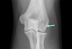
Radiographs of the elbow are typically normal in epicondylitis.[24][35] However, they may be useful to identify and evaluate calcifications or intra-articular pathology. About 22% to 25% of patients with epicondylitis may have calcifications within the soft tissues around the lateral epicondyle.[4][Figure caption and citation for the preceding image starts]: AP radiograph of elbow with lateral calcification from chronic lateral epicondylitisFrom the collection of Daniel J. Solomon, Naval Medical Center San Diego, CA; used with permission [Citation ends].
Computed tomography (CT), magnetic resonance imaging (MRI), ultrasound, and electromyographic studies are only performed if the history and physical examination findings do not clearly confirm the diagnosis, or if the patient has been refractory to conservative treatment.
CT scan or MRI of the elbow can be performed to evaluate the intra-articular anatomy, with particular regard to the presence of loose bodies, osteoarthritis, extra-articular pathology, or ligament damage, or the presence of a mass lesion.[12][39][40][41] MRI may be preferable to CT scan as it is able to demonstrate the quality of the tendon. Increased signal within the tendon of the extensor carpi radialis brevis or pronator may be seen in patients with lateral epicondylitis on MRI. [Figure caption and citation for the preceding image starts]: Coronal MRI and axial MRI in the same patient, showing high signal in extensor carpi radialis brevisFrom the collection of Daniel J. Solomon, Naval Medical Center San Diego, CA; used with permission [Citation ends].

Cervical MRI should be considered for patients who do not have a conclusive history and physical examination for epicondylitis, to investigate cervical radiculopathy as a cause for symptoms.[4][42][43]
Electromyogram and nerve conduction studies are performed if there is a sensory deficit or in the presence of muscle weakness not thought to be secondary to pain.
Ultrasound of the common extensor mass.[44] Accuracy may be dependent on a number of variables.[45] Operators should be qualified practitioners in detection of abnormal musculoskeletal ultrasound findings.[44]
Use of this content is subject to our disclaimer