Approach
Historic and physical examination findings are usually sufficient to make the diagnosis of wrinkles. Digital macrophotography, in vivo confocal microscopy, negative silicone rubber replicas, and histological examination of wrinkles are rarely indicated or necessary.
Clinical history
The age of the patient should be noted and a thorough social history taken. Information on the occupation, hobbies, and recreational activities of the patient should be sought, with particular attention to the degree of sun exposure received and the sun protection measures undertaken. The smoking status and dietary habits of the patient should also be elicited.
Physical examination
Wrinkles may begin at a young age, but are particularly evident in those over 40 years of age. They usually begin as fine lines, progressing to deep creases and folds with age, and are most noticeable in skin on exposed areas of the body (i.e., face, neck, dorsum of hands).
The face should be observed at rest and during animation. It should be noted if the wrinkles are obvious on motion or at rest, and whether they are superficial or deep, few or multiple, localised or extensive.[30]
By the fourth decade, wrinkles mostly appear with animation of the face; by the fifth decade, expressive lines result in permanent creases; and by the sixth to seventh decades, the whole facial skin may be covered with wrinkles. Gravitational lines are wrinkles in the form of sagging folds, which become more obvious between the ages of 40 and 50 years, and occur throughout the face and neck.[17][Figure caption and citation for the preceding image starts]: 30-year-old woman with mild wrinkles showing few wrinkles at restFrom the personal collection of Dr Dimitrios Dionyssiou; used with permission [Citation ends].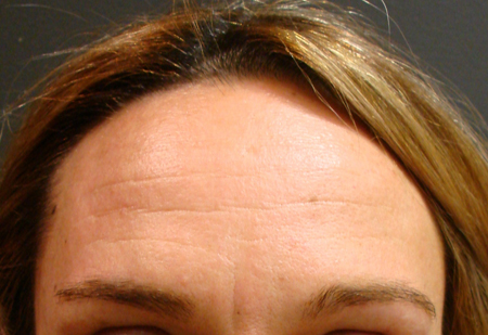 [Figure caption and citation for the preceding image starts]: 30-year-old woman with mild wrinkles showing moderate wrinkles on major facial activityFrom the personal collection of Dr Dimitrios Dionyssiou; used with permission [Citation ends].
 [Figure caption and citation for the preceding image starts]: 50-year-old woman with photo-aged skin showing advanced wrinkles, discoloration of the skin, actinic keratoses, and dynamic forces affecting the skinFrom the personal collection of Dr Dimitrios Dionyssiou; used with permission [Citation ends].
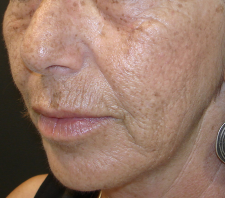 [Figure caption and citation for the preceding image starts]: 80-year-old woman with severe wrinkles, photoageing, and gravitational forces affecting the skinFrom the personal collection of Dr Dimitrios Dionyssiou; used with permission [Citation ends].
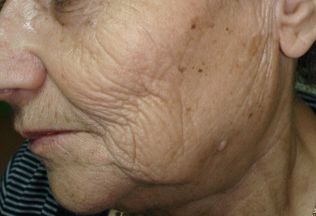
The site and distribution of the wrinkles should also be noted.[31] Wrinkles commonly manifest as horizontal forehead lines, glabellar lines between the eyebrows, lateral canthal lines ('crow's feet'), nasolabial folds, upper lip lines, lower radial lip lines, vertical lines at the corner of the mouth that run downward to the chin ('marionette lines'), a labiomental crease, cheek lines, and horizontal neck folds, as well as lines on the lower eyelids, on the dorsum of the hand and wrist, and in the preauricular area.
Repetitious hyperdynamic contractions of muscles responsible for facial expression pull directly on the overlying tissues, causing the skin to fold and develop dynamic lines. Among the first lines to develop (by the third decade) are the horizontal forehead creases, caused by contractions of the frontalis muscle, and lateral canthal or 'crow's feet' lines, caused by contractions of the orbicularis oculi muscle. Glabellar frown lines develop at variable ages as a result of the pull of the procerus and corrugator muscles.[5] Chronic contraction of the orbicularis oris muscle induces folds and lines around the mouth, perpendicular to the vermilion border, which become more noticeable in the fifth decade. Forceful contraction of the zygomaticus muscles contributes to the formation of the nasolabial folds.
Wrinkles resulting from intrinsic ageing are deep and often accompanied by skin laxity. Those due to sun exposure (photoageing or photodamage) are usually fine and are often accompanied by other signs such as dyschromia (liver spots, lentigines), dermal elastosis (advanced skin laxity), erythema, telangiectasia, textural changes (leathery appearance), and actinic keratoses.[32][33][34]Old, photoprotected skin may have increased laxity and fold accentuation, but it lacks signs of sun damage.[35]
Specialised investigations
The diagnosis of wrinkles is clinical, and therefore, further investigations are rarely required.
Macrophotography is a quality standardised form of high-resolution digital photography that demonstrates facial wrinkles with accuracy. It should be undertaken before any treatment and provides accurate pre- and post-treatment images.[19]
Skin biopsy with histological examination should only be performed to rule out other entities in the differential diagnosis.[36] The epidermis thins and atrophies with age. Desmoplakins outlining the cellular contours of keratinocytes are confirmatory. Under wrinkles, collagen (types IV and VII) is decreased in the dermoepidermal junction, and oxytalan fibres are decreased. There is deposition of abnormal elastotic tissue in the dermis, with interruption under wrinkles. Along with collagen atrophy, this magnifies the wrinkle depth. Chondroitin sulphates in the papillary dermis are decreased. Glycosaminoglycans are altered with asymmetric variation at the wrinkle edge. There is atrophy of the hypodermis with thickening of the fibrous lines. This makes the wrinkle depth more pronounced.
A mechanical or optical profilometer and an image-analysing computer are used to measure wrinkle depth. These measurements are confined to superficial wrinkles only, not facial furrows or folds.[31] Negative silicone rubber replicas of facial wrinkles are converted into hard, positive resin impressions and analysed directly using a simple light microscope.
In vivo confocal microscopy provides quantitative data on short- and long-term photodamage to the skin.[37][38] This technique is not used in clinical practice, and is used only for research purposes.
Use of this content is subject to our disclaimer