Oesophageal varices are a direct consequence of portal hypertension as a progressive complication of cirrhosis.
The development of bleeding carries significant morbidity and mortality.
Non-selective beta-blockers and/or endoscopic ligation can prevent the development of variceal bleeding.
Acute haemorrhage can be managed with resuscitation, terlipressin or a somatostatin analogue (e.g., octreotide), and endoscopic band ligation. Additional management includes prophylactic antibiotics and in some patients trans-jugular intrahepatic shunt therapy.
Diagnosis and surveillance by endoscopy is an important aspect of management.
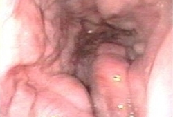
Oesophageal varices are dilated collateral blood vessels that develop as a complication of portal hypertension, usually in the setting of cirrhosis. They can be seen on endoscopy. In the US and Europe the major cause of cirrhosis is alcoholic liver disease. Worldwide, hepatitis B virus infection and hepatitis C virus infection are the major causes of cirrhosis.[1]Alter MJ. Epidemiology of hepatitis B in Europe and worldwide. J Hepatol. 2003;39 Suppl 1:S64-9.
https://www.journal-of-hepatology.eu/article/S0168-8278(03)00141-7/fulltext
http://www.ncbi.nlm.nih.gov/pubmed/14708680?tool=bestpractice.com
[2]Kim WR, Brown RS Jr, Terrault NA, et al. Burden of liver disease in the United States: summary of a workshop. Hepatology. 2002 Jul;36(1):227-42.
http://www.ncbi.nlm.nih.gov/pubmed/12085369?tool=bestpractice.com
Once cirrhosis has developed, increasing hepatic vein pressure gradient and deteriorating liver function may result in the formation of oesophageal varices.
Rupture of oesophageal varices can cause life-threatening bleeding. The most important predictor of variceal haemorrhage is the size of varices, with the highest risk of first haemorrhage occurring in patients with large varices (15% per year).[3]North Italian Endoscopic Club for the Study and Treatment of Esophageal Varices. Prediction of the first variceal hemorrhage in patients with cirrhosis of the liver and esophageal varices. A prospective multicenter study. N Engl J Med. 1988 Oct 13;319(15):983-9.
http://www.ncbi.nlm.nih.gov/pubmed/3262200?tool=bestpractice.com
[4]Garcia-Tsao G, Bosch J. Management of varices and variceal hemorrhage in cirrhosis. N Engl J Med. 2010 Mar 4;362(9):823-32.
http://www.ncbi.nlm.nih.gov/pubmed/20200386?tool=bestpractice.com
[5]Tripathi D, Stanley AJ, Hayes PC, et al. UK guidelines on the management of variceal haemorrhage in cirrhotic patients. Gut. 2015 Nov;64(11):1680-704.
https://gut.bmj.com/content/64/11/1680.long
http://www.ncbi.nlm.nih.gov/pubmed/25887380?tool=bestpractice.com
Other important predictors of haemorrhage are decompensated cirrhosis (Child-Pugh B/C) and the endoscopic finding of red wale marks.[3]North Italian Endoscopic Club for the Study and Treatment of Esophageal Varices. Prediction of the first variceal hemorrhage in patients with cirrhosis of the liver and esophageal varices. A prospective multicenter study. N Engl J Med. 1988 Oct 13;319(15):983-9.
http://www.ncbi.nlm.nih.gov/pubmed/3262200?tool=bestpractice.com
[5]Tripathi D, Stanley AJ, Hayes PC, et al. UK guidelines on the management of variceal haemorrhage in cirrhotic patients. Gut. 2015 Nov;64(11):1680-704.
https://gut.bmj.com/content/64/11/1680.long
http://www.ncbi.nlm.nih.gov/pubmed/25887380?tool=bestpractice.com
[6]Garcia-Tsao G, Abraldes JG, Berzigotti A, et al. Portal hypertensive bleeding in cirrhosis: risk stratification, diagnosis, and management: 2016 practice guidance by the American Association for the Study of Liver Diseases. Hepatology. 2017 Jan;65(1):310-35.
https://aasldpubs.onlinelibrary.wiley.com/doi/full/10.1002/hep.28906
http://www.ncbi.nlm.nih.gov/pubmed/27786365?tool=bestpractice.com
This topic covers oesophageal varices in adults.