Recommendations
Key Recommendations
Follow your local protocol to quickly identify and manage patients with suspected acute upper gastrointestinal (GI) bleeding; patients typically present with haematemesis and/or melaena.
Acute upper GI bleeding is a medical emergency requiring rapid intervention. In the UK, the British Society of Gastroenterology has produced a care bundle for the early clinical management of these patients.[35]
Suspect oesophageal variceal bleeding in a patient with upper GI bleeding who presents with:
Signs or symptoms of liver failure or decompensated cirrhosis including:[36][37]
Jaundice
Ascites
Hepatic encephalopathy
Physical signs of chronic liver disease (particularly cirrhosis) such as splenomegaly, spider angioma, and ascites
Deranged liver function tests and/or heavy prolonged alcohol misuse (patients who may have unrecognised chronic liver disease).
Admit any patient with suspected acute variceal bleeding to a high-dependency or intensive care unit.[5] Haemorrhage from oesophageal varices is a life-threatening emergency with a high mortality rate.[36][37] Prompt diagnosis (and intervention) is essential to improve the clinical outcome.
Perform a risk assessment. If Glasgow-Blatchford score ≤1, consider managing the patient as an outpatient if safe and appropriate to do so.[35][38]
Refer all patients with suspected acute upper GI bleeding for upper GI endoscopy.[35][38][39]
If the patient is unstable with severe acute upper GI bleeding, do this urgently, immediately after resuscitation.[35][39]
For all other patients with upper GI bleeding, do this within 24 hours of admission.[35][39]
Intubate the patient before endoscopy if there is haematemesis, or there is a perceived risk of a haemodynamically unstable patient having blood in the stomach, or if the patient presents with an altered mental status (i.e., confusion).[5]
Haemorrhage from oesophageal varices is a life-threatening emergency with a high mortality rate.[36][37] Prompt diagnosis (and intervention) is essential to improve the clinical outcome.
Oesophageal varices are a progressive complication of cirrhosis and a direct consequence of portal hypertension. See our topic Cirrhosis.
Suspect oesophageal variceal bleeding in a patient with upper gastrointestinal (GI) bleeding who presents with:
Signs or symptoms of liver failure or decompensated cirrhosis including:[37]
Jaundice
Ascites
Hepatic encephalopathy
Physical signs of chronic liver disease (particularly cirrhosis) such as splenomegaly, spider angioma, and ascites
Deranged liver function tests and/or heavy prolonged alcohol misuse (patients who may have unrecognised chronic liver disease).
Patients with active variceal bleeding typically present with haematemesis and/or melaena.
Patients with haematemesis tend to have more severe bleeds than those with only melaena.[40]
Patients who are haemodynamically unstable commonly present with haematochezia.
Admit any patient with suspected acute variceal bleeding to a high-dependency unit.[5]
When assessing a patient with acute upper GI bleeding, consider other causes, such as peptic ulceration, gastric erosions, and use of medication such as non-steroidal anti-inflammatory drugs. See the Differentials section.
Varices are the cause of haemorrhage in about 10% of patients admitted with acute GI bleeding in the UK.[5][19]
Practical tip
Assess any patient presenting with an unexplained postural hypotension or syncope for an unrecognised GI haemorrhage.
Bear in mind that varices may be asymptomatic in patients without haemorrhage. Evaluate for signs and symptoms of liver disease and extent of cirrhosis to help stratify the risk for bleeding.
In the community
Refer any patient who presents with suspected acute upper GI bleeding (i.e., haematemesis and/or melaena) urgently to hospital. Patients with suspected acute variceal bleeding should be admitted to a high-dependency unit, as variceal bleeding is unpredictable and associated with high mortality. A specialist monitored bed for gastrointestinal bleeding may also be appropriate.[5]
Take a full history. Ask about:
History of alcohol misuse
Excessive chronic alcohol consumption is a risk factor for development of alcoholic hepatitis and alcoholic cirrhosis
History of intravenous drug use
People who inject drugs are at risk of contracting hepatitis B and hepatitis C, which can cause cirrhosis
Cirrhosis
About 50% of patients with cirrhosis have gastro-oesophageal varices[41]
Common precipitants of decompensation of cirrhosis include:[37]
Gastrointestinal bleeding (variceal and non-variceal)
Infection/sepsis (spontaneous bacterial peritonitis, urine, chest, cholangitis, etc.)
Alcohol hepatitis
Acute portal vein thrombosis
Development of hepatocellular carcinoma
Drugs (alcohol, opioids, non-steroidal anti-inflammatory drugs, etc.)
Ischaemic liver injury (sepsis or hypotension)
Dehydration
Constipation
Viral hepatitis
Chronic hepatitis B or C infection may lead to cirrhosis and oesophageal varices. The incidence of oesophageal varices is increased when viral hepatitis is combined with alcohol misuse
HIV co-infection
May accelerate liver disease in patients with chronic viral hepatitis
Haemochromatosis, Wilson's disease, and other recognised causes of portal hypertension, such as Budd-Chiari syndrome, myeloproliferative disorders, and sarcoidosis.
Practical tip
Watch for typical alcohol and/or drug withdrawal symptoms after admission.[5] See our topics Alcohol withdrawal and Opioid use disorder.
The risk of variceal bleeding increases with:[3][4][5][6]
The size of varices
The presence of red wale marks (defined as longitudinal dilated venules resembling whip marks on the variceal surface)
The severity of liver disease.
Look for signs of:
Liver failure or decompensated cirrhosis including:[37]
Jaundice
Increasing ascites
Hepatic encephalopathy
Chronic liver disease (particularly cirrhosis) including:
Ascites
Evidence of decompensated cirrhosis, which is an important factor associated with development and progression of varices
Jaundice
May indicate advanced liver disease
Splenomegaly
Patients with splenomegaly often have thrombocytopenia and anaemia, owing to pooling of blood and sequestration of blood components in the spleen in the setting of portal hypertension
Spider angioma, bruising, petechiae
Multiple spider angiomas are a feature of chronic liver disease and cirrhosis. The number and size correlate with the severity of chronic liver disease. Patients with numerous large angiomas are at increased risk of variceal haemorrhage
Caput medusa
Abdominal wall vascular collaterals are an important clinical sign and consequence of severe portal hypertension
Encephalopathy (i.e., confusion, coma)
Evidence of decompensated cirrhosis.
Active variceal bleeding typically presents with haematemesis and/or melaena.
Patients with haematemesis tend to have more severe bleeds than those with only melaena.[40] Patients who are haemodynamically unstable commonly present with haematochezia.
Practical tip
In patients with shock and no apparent cause, perform a rectal examination early to avoid missing melaena.
On presentation or admission, perform urgent observations on patients with suspected acute upper gastrointestinal (GI) bleeding using a validated early warning score.[35] The British Society of Gastroenterology (BSG) recommends the National Early Warning Score (NEWS). Use NEWS2, or a similarly validated early warning score, to help determine how quickly the patient needs an endoscopy.[35] Royal College of Physicians: National Early Warning Score (NEWS) 2 Opens in new window
Use prognostic risk assessment scores, as recommended by the UK National Institute for Health and Care Excellence (NICE), the BSG and the European Society of Gastrointestinal Endoscopy (ESGE) in all patients with acute upper GI bleeding.[35][38][39] Use the scores to help decide which patients need urgent endoscopy (within the next 12 hours), which patients are stable enough to be admitted and have non-urgent endoscopy (within 24 hours), and which patients can be discharged from hospital.[42][43]
Use the Glasgow-Blatchford score at first assessment to identify patients with low risk bleeds who can be safely discharged without undergoing endoscopy.[35][38][39] [ Blatchford Score for Gastrointestinal Bleeding Opens in new window ]
If Glasgow-Blatchford score ≤ 1, consider managing the patient as an outpatient if safe and appropriate to do so. This approach is supported by latest evidence and in line with recommendations from the BSG and ESGE.[35][38]
Bear in mind that recommendations on discharge from NICE (early discharge for patients with a pre-endoscopy Blatchford score of 0) are based on older evidence and therefore do not reflect current practice.[39]
Use the full Rockall score after endoscopy as recommended by NICE.[39] [ Rockall Score for Upper Gastrointestinal Bleeding Opens in new window ]
A score of ≤2 is associated with a low risk of further bleeding or death.
Calculate the Child-Pugh score, based on biochemical and clinical findings, and determine the Child-Pugh class in patients with cirrhosis. [ Child Pugh classification for severity of liver disease (SI units) Opens in new window ]
Refer all patients with suspected acute upper gastrointestinal (GI) bleeding for upper GI endoscopy (i.e., gastroscopy).[35][38][39]
If the patient is unstable with severe acute upper GI bleeding, do this urgently, immediately after resuscitation.[35][39]
For all other patients with upper GI bleeding, do this within 24 hours of admission.[35] [39]
Intubate the patient before endoscopy if there is haematemesis, or there is a perceived risk of a haemodynamically unstable patient having blood in the stomach, or if patient presents with an altered mental status (i.e., confusion).[5] Make sure the patient is appropriately resuscitated before undergoing endoscopy.
Practical tip
If the patient has been intubated, they will need to be managed in an intensive care or high-dependency unit until extubation.[5]
Gastroscopy is the gold standard technique for the diagnosis of varices and the assessment of variceal size.[5][6][7][44][45][46][47]
Variceal size is the most important predictor of haemorrhage, with the highest risk of first haemorrhage occurring in patients with large varices (15% per year).[3][4] Large varices occupy more than one third of the oesophageal lumen.[3] See the Criteria section.
The endoscopic finding of red wale marks (defined as longitudinal dilated venules resembling whip marks on the variceal surface) and the severity of liver disease are also predictive factors.[3][5][6]
[Figure caption and citation for the preceding image starts]: Large oesophageal varicesFrom the personal collection of Gennaro D'Amico, MD [Citation ends].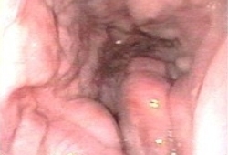 [Figure caption and citation for the preceding image starts]: Large oesophageal varicesFrom the personal collection of Gennaro D'Amico, MD [Citation ends].
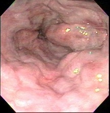
More info: Contraindications for upper GI endoscopy
Absolute contraindications for an upper GI endoscopy include:
Severe hypotension/shock
Acute perforation
Acute myocardial infarction
Peritonitis.
Relative contraindications for an upper GI endoscopy include:
Uncooperative patient
Coma (except those already intubated)
Cardiac arrhythmias
Myocardial ischaemia (recent event).
Capsule endoscopy may have a role in screening of varices in patients who are not candidates for gastroscopy, in patients who refuse gastroscopy, or if gastroscopy is not available.[5][48] However, sensitivity of capsule endoscopy is not sufficient to replace gastroscopy as an initial exploration.[5][48][49]
In patients with confirmed variceal bleeding, order a liver ultrasound to screen for portal vein thrombosis (PVT) and hepatocellular carcinoma (HCC). If ultrasound identifies PVT/HCC, seek advice on management from a gastroenterologist.
Request a full blood count, electrolytes, liver function tests, renal function tests, and coagulation profile.[5]
If the patient’s history suggests viral hepatitis as a cause of cirrhosis, also request a hepatitis B surface antigen (HBsAg) and anti-hepatitis C virus immunoglobulin G (anti-HCV IgG).
Send blood for typing and cross-matching.
Patients with upper gastrointestinal bleeding can experience rapid clinical deterioration. Blood products may become necessary.
Use of this content is subject to our disclaimer