Images and videos
Images

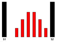


Aortic stenosis
The systolic crescendo-decrescendo murmur of aortic stenosis
From the collection of David Liff, MD, Emory University Hospital; used with permission
See this image in context in the following section/s:
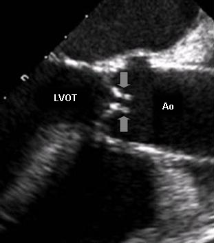
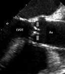
Aortic stenosis
Transoesophageal echocardiogram showing the left ventricle outflow tract (LVOT), the aorta (Ao), and nearly immobile leaflets (arrows) of a severely stenotic aortic valve
From the collection of David Liff, MD, Emory University Hospital; used with permission
See this image in context in the following section/s:

Aortic stenosis
Balloon valvuloplasty fluoroscopy film that demonstrates valvuloplasty balloon inflated across a calcified aortic valve
From the collection of David Liff, MD, Emory University Hospital; used with permission
See this image in context in the following section/s:
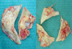
Aortic stenosis
Bicuspid and trileaflet aortic valves with severe calcification following surgical excision
From the collection of David Liff, MD, Emory University Hospital; used with permission
See this image in context in the following section/s:
Aortic stenosis
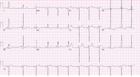
ECG showing changes associated with left ventricular hypertrophy
From the collection of Melanie Everitt MD, Heart Failure & Transplantation Program, Primary Children's Medical Center, Salt Lake City, UT; used with permission
See this image in context in the following section/s:
Videos
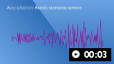 Aortic stenosis (severe)
Auscultation sounds: Aortic stenosis (severe)
 How to perform an ECG animated demonstration
How to record an ECG. Demonstrates placement of chest and limb electrodes.
Use of this content is subject to our disclaimer