Images and videos
Images


Merkel cell carcinoma
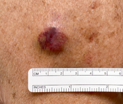
A 2 cm red, nodular MCC with varied vessel patterns on the right upper arm of an 80-year-old man
From the collection of Dr Kelly Harms and Dr Alison Lee, University of Michigan; used with patient consent
See this image in context in the following section/s:
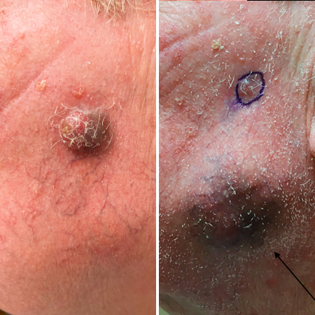
Merkel cell carcinoma
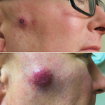
MCC presenting as a well-defined, rapidly growing, asymptomatic, brightly violaceous nodule on the left cheek of a 71-year-old man. The patient developed a golf-ball sized subcutaneous mass in the left parotid (original biopsy site marked with blue ink, subcutaneous metastasis marked with arrow). Fine needle aspiration of the mass in the left parotid gland confirmed MCC
From the collection of Dr Kelly Harms and Dr Alison Lee, University of Michigan; used with patient consent
See this image in context in the following section/s:

Merkel cell carcinoma
A rapidly-growing MCC on the right cheek of a 64-year-old man, which started as a small erythematous cyst-like papule then developed within a few months into a larger, violaceous nodule with associated pruritus
From the collection of Dr Kelly Harms and Dr Alison Lee, University of Michigan; used with patient consent
See this image in context in the following section/s:
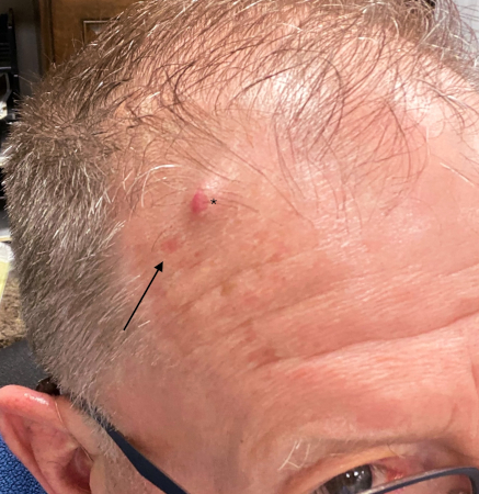
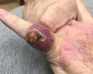
Merkel cell carcinoma
Primary MCC on the right frontal scalp (asterisk) of a 73-year-old man, with a biopsy scar of an in-transit metastasis (arrow). The primary tumor initially presented as an asymptomatic, fast-growing subcutaneous nodule that was misdiagnosed as an epidermoid cyst
From the collection of Dr Kelly Harms and Dr Alison Lee, University of Michigan; used with patient consent
See this image in context in the following section/s:

Merkel cell carcinoma
Histological features of MCC from biopsy of a primary tumour. Image B shows small round blue cells and image C shows characteristic nuclei, finely granular and dusty 'salt and pepper' chromatin, and abundant mitotic figures
Mauzo SH et al. J Clin Pathol 2016; 69: 382-90; used with permission
See this image in context in the following section/s:
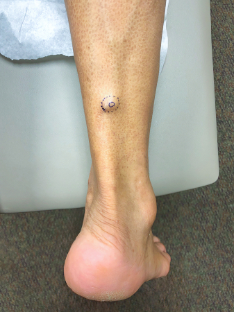
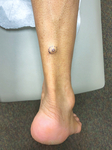
Merkel cell carcinoma
MCC on the right posterior ankle of a 66-year-old African-American man, which presented as a subtle 1.5 cm subcutaneous nodule without overlying epidermal changes. This can often be confused with benign entities such as an epidermal cyst or a lipoma
From the collection of Dr Kelly Harms and Dr Alison Lee, University of Michigan; used with patient consent
See this image in context in the following section/s:

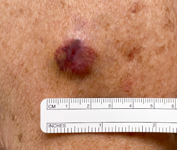
Merkel cell carcinoma
A 2x3 cm MCC lesion in a white man aged in his late 60s who had a history of taking immunosuppressive medication for psoriasis. The nodule was violaceous with central ulceration, tan crusting, and serous drainage. Note that ulceration is unusual in MCC
Tomtschik J et al. BMJ Case Reports CP 2022; 15: e249288; used with permission
See this image in context in the following section/s:
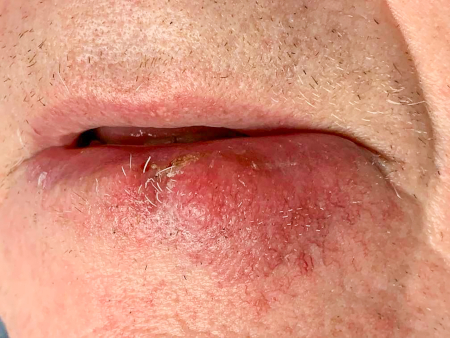
Merkel cell carcinoma
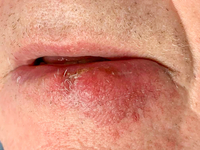
MCC on the left lower lip of a 68-year-old man. The asymptomatic tumour is an ill-defined reddish-pink, scaly plaque blurring the lower vermillion of the lip
From the collection of Dr Kelly Harms and Dr Alison Lee, University of Michigan; used with patient consent
See this image in context in the following section/s:

Merkel cell carcinoma
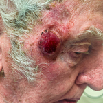
A primary MCC tumour in an 87-year-old man who presented with a large, fast-growing, red, eroded nodule on the right temple
From the collection of Dr Kelly Harms and Dr Alison Lee, University of Michigan; used with patient consent
See this image in context in the following section/s:
Use of this content is subject to our disclaimer