Images and videos
Images


Hospital-acquired pneumonia (non COVID-19)
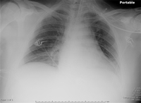
Increased opacification of the right perihilar region and superior segment of the right lower and upper lobes consistent with worsening aspiration pneumonia
From the collection of Dr Roy Hammond, MD; used with permission
See this image in context in the following section/s:

Hospital-acquired pneumonia (non COVID-19)
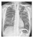
Chest x-ray showing left upper lobe cavitating pneumonia
From the collection of Dr Jonathan Bennett; used with permission
See this image in context in the following section/s:

Hospital-acquired pneumonia (non COVID-19)
CT scan showing bi-basilar opacities in a patient with HAP
Consent obtained at University of Louisville, Louisville, KY
See this image in context in the following section/s:

Hospital-acquired pneumonia (non COVID-19)
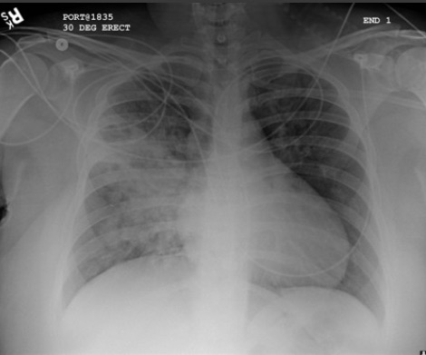
Portable chest x-ray of a patient with HAP. Note the obscured left hemidiaphragm due to a left lower lobe opacity and an obscured heart border due to a left upper lobe or lingular opacity
Consent obtained at University of Louisville, Louisville, KY
See this image in context in the following section/s:

Hospital-acquired pneumonia (non COVID-19)
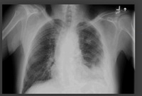
Left-sided pleural effusion
From the collection of Dr R Light; used with permission
See this image in context in the following section/s:
Use of this content is subject to our disclaimer