Images and videos
Images


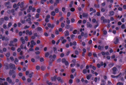
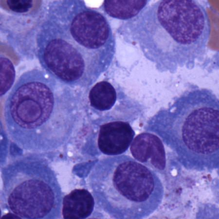
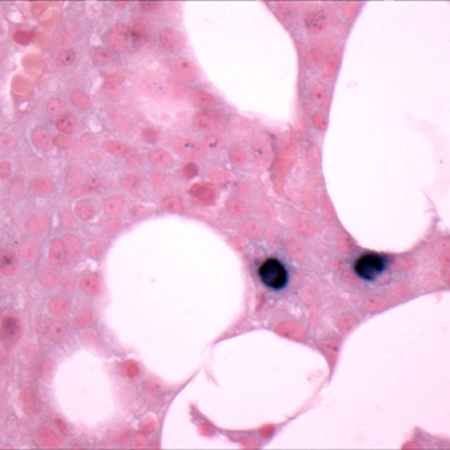
Multiple myeloma
Bone marrow biopsy after histochemical analysis for kappa light chain
Courtesy of Dr Robert Hasserjian, Hematopathology, Massachusetts General Hospital; used with permission
See this image in context in the following section/s:
Multiple myeloma
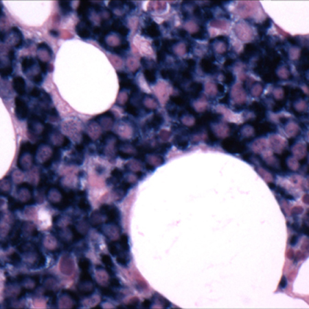
Bone marrow biopsy
Courtesy of Dr Robert Hasserjian, Hematopathology, Massachusetts General Hospital; used with permission
See this image in context in the following section/s:

Multiple myeloma
A: serum protein electrophoresis demonstrating a monoclonal immunoglobulin. B: serum and urine immunofixation electrophoresis (IFE) demonstrating a monoclonal immunoglobulin and free light chain (Bence Jones [BJ] protein)
Courtesy of Dr M Murali and the Clinical Immunology Laboratory, Massachusetts General Hospital; used with permission
See this image in context in the following section/s:
Multiple myeloma
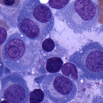
Aspirate showing plasma cell infiltrate
Courtesy of Dr Robert Hasserjian, Hematopathology, Massachusetts General Hospital; used with permission
See this image in context in the following section/s:
Multiple myeloma
A: serum protein electrophoresis (SPEP) of normal serum. B: SPEP of multiple myeloma serum showing a monoclonal immunoglobulin (M-protein) in the gamma region. C: densitometry tracing of normal serum (A) showing the 5 zones of the high resolution agarose electrophoresis. D: densitometry tracing of multiple myeloma serum (B) showing a monoclonal spike (M spike)
Courtesy of Dr M Murali and the Clinical Immunology Laboratory, Massachusetts General Hospital; used with permission
See this image in context in the following section/s:
Multiple myeloma
Bone marrow biopsy after histochemical analysis for lambda light chain
Courtesy of Dr Robert Hasserjian, Hematopathology, Massachusetts General Hospital; used with permission
See this image in context in the following section/s:
Use of this content is subject to our disclaimer