Investigations
1st investigations to order
ectoparasite preparation
Test
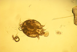
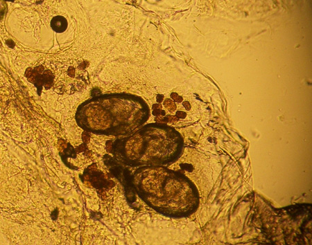
Should be performed on any patient suspected to have scabies, but should not delay treatment if there is a high index of clinical suspicion. Specificity is 100% if a mite is seen; sensitivity is generally <50% and depends on experience level of the clinician.[5][Figure caption and citation for the preceding image starts]: Scabies mite under 10× powerFrom the collection of Laura Ferris, MD, PhD [Citation ends]. [Figure caption and citation for the preceding image starts]: Eggs and stool under 10× powerFrom the collection of Laura Ferris, MD, PhD [Citation ends].
Result
presence of mite, eggs, or faecal material of mites
Investigations to consider
skin biopsy
Test
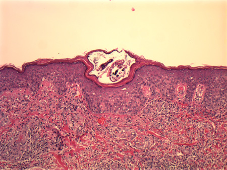
Skin biopsy may be done to look for an alternative diagnosis if ectoparasite is negative. However, it is rare to see a mite in the biopsy specimen, and the eosinophilic infiltrate seen in most biopsies is non-specific and not sufficient for diagnosis.[17][Figure caption and citation for the preceding image starts]: Histological section showing an adult Sarcoptes scabiei in its burrow in the stratum corneumFrom the collection of Pooja Khera, MD [Citation ends].
Result
presence of mite, eosinophilic infiltrate; rarely, eggs and mite faecal material are found
Emerging tests
epiluminescence light microscopy
Test
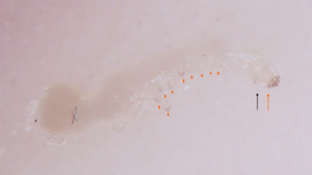
This is a technique noted to be 93% sensitive in the diagnosis of clinically suspected cases of scabies. It has the advantage of being inexpensive and non-invasive.[15] Has been shown to be an accurate method for diagnosis when performed by a properly trained practitioner.[21][Figure caption and citation for the preceding image starts]: Classic dermoscopic image of triangle or 'delta wing jet' sign of dense scabies head parts (long red arrow), relatively translucent scabies body (long black arrow), scabies eggs (short red arrows), and classic S-shaped burrowFox G. Diagnosis of scabies by dermoscopy. BMJ Case Rep. 2009;2009.pii: bcr06.2008.0279. [Citation ends].
Result
presence of the mites indicated by small, dark, triangular structures located at one end of the linear burrow
Use of this content is subject to our disclaimer