Images and videos
Images

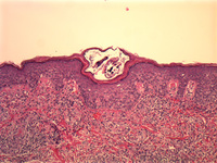
Scabies
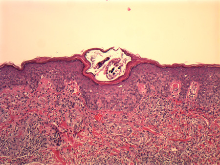
Histological section showing an adult Sarcoptes scabiei in its burrow in the stratum corneum
From the collection of Pooja Khera, MD
See this image in context in the following section/s:

Scabies
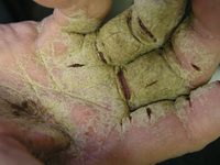
Hyperkeratotic palms in a patient with Norwegian scabies
Photograph courtesy of Joseph C. English, III, MD
See this image in context in the following section/s:
Scabies
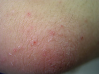
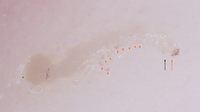
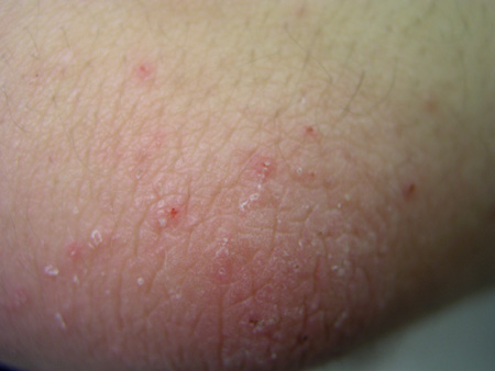
Characteristic linear burrows in skin
From the collection of Laura Ferris, MD, PhD
See this image in context in the following section/s:

Scabies
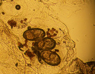
Eggs and stool under 10× power
From the collection of Laura Ferris, MD, PhD
See this image in context in the following section/s:

Scabies
Classic dermoscopic image of triangle or 'delta wing jet' sign of dense scabies head parts (long red arrow), relatively translucent scabies body (long black arrow), scabies eggs (short red arrows), and classic S-shaped burrow
Fox G. Diagnosis of scabies by dermoscopy. BMJ Case Rep. 2009;2009:bcr06.2008.0279.
See this image in context in the following section/s:
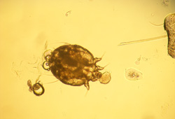
Scabies
Scabies mite under 10× power
From the collection of Laura Ferris, MD, PhD
See this image in context in the following section/s:
Scabies
Classic dermoscopic image of triangle or 'delta wing jet' sign of dense scabies head parts (long red arrow), relatively translucent scabies body (long black arrow), scabies eggs (short red arrows), and classic S-shaped burrow
Fox G. Diagnosis of scabies by dermoscopy. BMJ Case Rep. 2009;2009.pii: bcr06.2008.0279.
See this image in context in the following section/s:

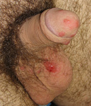
Scabies
Penile nodules, characteristic of scabies
Photograph courtesy of Joseph C. English, III, MD
See this image in context in the following section/s:
Use of this content is subject to our disclaimer