Differentials
Lymphoma
SIGNS / SYMPTOMS
History of persistently enlarged lymph nodes, constitutional or B symptoms (fevers, night sweats, and/or weight loss). Usually under age 35 or over age 65. Physical examination may reveal lymphadenopathy in one or more regions; hepatosplenomegaly may be present.
If there is involvement of the anterior mediastinum alone, it may be difficult to differentiate lymphoma from thymic malignancy radiographically.
INVESTIGATIONS
Chest CT scan usually shows a more multinodular, aggressive, or invasive mass, but not always. If there is peripheral lymphadenopathy, excision biopsies will reveal the type of lymphoma. If lymphoma involves the anterior mediastinum alone, core needle biopsy will be required to differentiate from thymic malignancy.
Malignant mediastinal germ cell tumour: seminoma
SIGNS / SYMPTOMS
Occurs in people aged 20 to 40 years, with male predominance.
Presenting symptoms depend on tumour location, growth rate, and size; they include chest pressure, hoarseness, chest wall pain, dysphagia, and dyspnoea.
INVESTIGATIONS
Chest CT scan usually shows homogeneous invasive malignant-appearing anterior mediastinal mass.
Serum alpha-fetoprotein normal; may have low-level elevation of serum human chorionic gonadotrophin (beta-hCG).
Malignant mediastinal germ cell tumour: non-seminoma
SIGNS / SYMPTOMS
Patient is typically a young male with significant symptoms of systemic and local invasion, such as fatigue, weight loss, fever, dyspnoea, and chest pain.
INVESTIGATIONS
Serum alpha-fetoprotein and human chorionic gonadotrophin (beta-hCG) usually elevated.
Chest CT scan usually shows a large, inhomogeneous invasive mass with areas of haemorrhage and necrosis; pleural effusion often present; may show lung metastases.
Benign mediastinal germ cell tumour
SIGNS / SYMPTOMS
Typically asymptomatic because of slow growth rate of the tumour.
If present, the most common symptoms are dyspnoea and substernal chest pain.
INVESTIGATIONS
On chest CT, mature (low-grade) teratoma has a pathognomonic CT appearance with cystic, fatty, and/or calcified components.
Substernal goitre
SIGNS / SYMPTOMS
Older patient usually without symptoms. Rarely, patients have upper-chest discomfort, dyspnoea, or hoarseness. Symptoms of hyperthyroidism (heat intolerance, weight loss) or hypothyroidism may be present.
A visible goitre is present in 80% to 90% of patients; substernal extension is suggested if the lower pole of the thyroid gland cannot be identified. Airway compression may present with stridor and prolonged inspiration/expiration. Signs of thyrotoxicosis, including weight loss, hypertension, and tachycardia, may be evident on physical examination. Tracheal deviation can occur if the goitre is asymmetrical. Pemberton's signs (neck vein distension and facial flushing when arms are held vertically above head [Pemberton's manoeuvre]) may be present.
INVESTIGATIONS
Chest CT shows anterior mediastinal mass nearly always contiguous with the thyroid, and tending to also involve the middle mediastinum/paratracheal space; mass often has low-density areas and calcifications; if goitre is suspected, iodinated contrast should be avoided (due to risk of iodine-induced hypothyroidism; Jod-Baselow effect).
Thyroid function tests may be abnormal.
Thymic hyperplasia
SIGNS / SYMPTOMS
Usually asymptomatic, but can be seen with myasthenia gravis, Graves' disease, burns; following viral illnesses; and as a rebound phenomenon (e.g., following chemotherapy or corticosteroid therapy, especially for Hodgkin's disease).
INVESTIGATIONS
Chest CT usually shows enlargement of the typical shape of the thymus (bilobed) without a dominant or discrete rounded mass. [Figure caption and citation for the preceding image starts]: CT scan of the chest showing a prominent thymic gland with bilobed appearance, consistent with thymic hyperplasiaFrom the collection of Cameron Wright, MD; used with permission [Citation ends].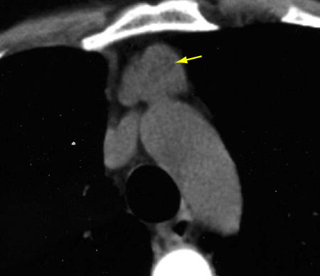
Chemical-shift MRI may be helpful in differentiating hyperplasia from tumour in problematical cases.
Thymic cyst
SIGNS / SYMPTOMS
Usually asymptomatic and an incidental finding. Some cysts can be large and heterogenous on imaging.
INVESTIGATIONS
MRI is helpful to distinguish between a cyst and solid tumour. No fluorine-18 fluorodeoxyglucose-avidity with PET scans.
Use of this content is subject to our disclaimer