Tests
1st tests to order
clinical exam
Test
It is not routinely recommended to obtain x-rays before consulting a spine specialist, as these will routinely be obtained upon the patient's presentation to the specialist.
Result
an asymmetric thoracic, thoracolumbar, or lumbar paraspinal prominence may be observed when patient bends forward
Tests to consider
standing PA x-rays of cervical, thoracic, and lumbar spine, and pelvis
Test
Should include visualization of the entire spine (i.e., from the cervicothoracic junction to the hips), as well as the pelvis, on a single film.
Each vertebra should be counted as well as each rib and pedicle, bilaterally, in order to identify failures of segmentation or formation that may be the cause of the deformity.
Allow evaluation of the iliac apophysis (Risser sign) and acetabular tri-radiate cartilage in order to calculate bone age and estimate remaining growth potential.[23][24][25][27][36] There is no evidence of an apophysis in the most immature patients, while in mature patients it is fully developed. Closure of the triradiate cartilage coincides with the end of the peak adolescent growth spurt.[27]
Allow determination of the extent of coronal decompensation by comparing the distance between the C7 plumb line (C7PL) and the central sacral vertical line (CSVL).
The Cobb angle should be measured for all major and minor curves. An angle >10° establishes a diagnosis of scoliosis.[Figure caption and citation for the preceding image starts]: Posteroanterior scoliosis radiograph of a 13-year-old girl with a 49° right thoracic curvature with apex at the T9-T10 disc spaceFrom the collection of Stuart Weinstein, MD, University of Iowa; used with permission [Citation ends].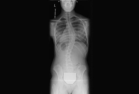
Result
positive finding: coronal plane spinal curvature >10°
standing lateral x-rays of cervical, thoracic, and lumbar spine, and pelvis
Test
Should include visualization of the entire spine (i.e., from the cervicothoracic junction to the hips), as well as the pelvis, on a single film.
The sagittal balance should be determined by measuring the distance from the C7PL to the posterior superior corner of the S1 vertebral body (the C7PL should pass through the posterior superior corner of S1 on a standing lateral projection).
It is also possible to measure the degree of thoracic kyphosis and lumbar lordosis from the standing lateral radiograph.
Positive findings are subtler on lateral than on PA films.[Figure caption and citation for the preceding image starts]: Lateral scoliosis radiograph of a 13-year-old girl with a 49° right thoracic curvatureFrom the collection of Stuart Weinstein, MD, University of Iowa; used with permission [Citation ends].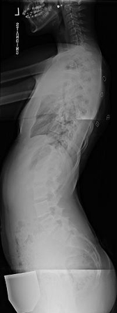
Result
positive finding: hypo- or hyperkyphosis of the thoracic spine
MRI of cervical, thoracic, lumbar, and sacral spine, and brainstem
Test
Do not order MRI for scoliosis in a child until all appropriate clinical and plain radiographic exams have been completed.[39]
Only recommended in patients with an atypical presentation or abnormality on neurologic exam to rule out intraspinal pathology (e.g., syringomyelia, Arnold-Chiari malformation, intramedullary tumor/mass) as the inciting cause of the spinal deformity.
Should be undertaken with and without gadolinium contrast.
Result
normal
Use of this content is subject to our disclaimer