History and exam
Key diagnostic factors
common
age >10 years
In order to be diagnosed with adolescent idiopathic scoliosis (AIS), the patient must be at least 10 years of age.
6 to 12 months post puberty
Menarche in girls or development of axillary and facial hair in boys should be assessed to estimate the onset of the peak adolescent growth spurt, as these features occur 6 to 12 months after this period.
postural asymmetry
Asymmetry of posture may be noted during activities, while looking in the mirror, during daily hygiene, or while dressing.
absent or minimal pain
Back pain is usually minimal or absent at presentation. Significant pain at presentation should warrant a careful evaluation for other causes of the spinal deformity.
absence of neurologic symptoms with normal neurologic exam
Patients with AIS should not have abnormal neurologic symptoms at presentation and the neurologic examination is normal. Even subtle abnormalities, such as sensation changes or motor weakness, warrant advanced neurologic imaging with MRI.
paraspinal prominences on forward bending
Asymmetric thoracic, thoracolumbar, or lumbar paraspinal prominences result from abnormal vertebral rotation as well as a combination of abnormal spinal curvature in the coronal and sagittal planes.
The presence of such a prominence is the hallmark exam finding that leads to a suspicion of scoliosis during screening evaluation with Adams forward bend test.
A positive result on Adams forward bend test is observation of an asymmetric paraspinal prominence. The presence of an asymmetric scapular prominence may suggest an upper thoracic curve.[33]
This key sign may be recognized by the patient before or after the diagnosis is made.
scoliometer measurement of >5° at paraspinal prominence
This measurement allows quantification of the paraspinal prominences (which result from abnormal vertebral rotation as well as a combination of abnormal spinal curvature in the coronal and sagittal planes) found by Adams forward bend test.
A positive result is one of >5° at any paraspinal prominence (thoracic or lumbar).
Although scoliometer measurements do not exactly represent Cobb angle measurements from plain film radiographs, the 2 values may correlate with one another. In general, scoliometer measurements of 5° and 7° correspond to a measured Cobb angle of approximately 10° and 20°, respectively.[34][35]
This correlation makes scoliometer measurements useful, as they represent commonly agreed-upon cut-off points used to direct treatment decisions.[Figure caption and citation for the preceding image starts]: Scoliometer measurement of a right thoracic prominenceWeinstein SL, et al. Adolescent idiopathic scoliosis. Lancet. 2008;371:1527-1537. Used with permission [Citation ends].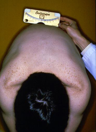
symmetric abdominal reflexes
Abdominal reflexes are assessed to rule out intramedullary lesions of upper motor neurons.
Patients with AIS should have normal abdominal reflexes. The umbilicus should deviate toward the abdominal quadrant being stimulated during the examination. Asymmetry or absence of this reflex is suggestive of underlying neuropathology.
Other diagnostic factors
common
shoulder asymmetry
Results from abnormal spinal curvature in the coronal plane.
In the majority of cases it is present on the convex side of the major structural curve, most commonly with elevation of the right shoulder in a right main thoracic structural curve.
This may be recognized by the patient before or after the diagnosis is made.
waist-line asymmetry
May signify truncal decompensation (severe, clinically apparent deformity in which the trunk is no longer centered over the pelvis, resulting from coronal imbalance in severe curves) from a spinal deformity. May also be present in patients with leg-length discrepancy.
This may be recognized by the patient before or after the diagnosis is made.
thoracic wall or breast asymmetry
This is a result of abnormal vertebral rotation that alters rib orientation, resulting in a change in shape between the 2 hemithoraces.
This may be recognized by the patient before or after the diagnosis is made.
uncommon
normal gag reflex
Patients with AIS have a normal gag reflex. An abnormal gag reflex may be an abnormality involving the hindbrain, such as in Arnold-Chiari malformation.
truncal decompensation
In more severe curves, coronal imbalance can develop, resulting in a severe, clinically apparent deformity in which the trunk is no longer centered over the pelvis.
Risk factors
strong
positive family history
The prevalence of scoliosis in the daughters of women with adolescent idiopathic scoliosis (AIS) is almost 30%.[13] Additionally, twin concordance studies have shown a prevalence of 73% to 92% in monozygotic twins and 36% to 63% in dizygotic twins.[13][14][15]
Despite these observations, specific genetic abnormalities have not been consistently shown.[16][17][18][19] The true inheritance pattern of AIS is likely to be multifactorial.
peak adolescent growth spurt
Scoliosis curve development and progression have been shown to correlate closely with the period of peak skeletal growth during adolescence.
A patient’s remaining growth potential is estimated using objective clinical data, such as pubertal status and bone age, commonly measured using the Risser sign and Sanders Maturity Score. The Risser sign assesses bone age through the evaluation of the developing iliac apophysis.[23]
The iliac apophysis develops from lateral to medial on a coronal view, with the most immature patients having no evidence of an apophysis and the most mature having a fully developed and fused apophysis that extends to the sacroiliac joint. The risk for curve progression in patients with a Risser sign of 1 or less can be as high as 70%. In a patient with a Risser sign of 3, the risk of progression is approximately 10%.[24][25]
The Sanders maturity score (skeletal maturity scoring system) has been found to be more reliable than the Risser sign as a clinical maturity indicator.[26] This system uses radiographs of the patient’s hand to evaluate the radiographic appearance of epiphyses of the distal part of the radius and ulna, along with metacarpals and phalanges. Patients at the greatest risk for significant curve progression were those with curves >20° at stage 2 and >30° at stage 3.[26]
Another method is evaluation of the acetabular tri-radiate cartilage on PA pelvis x-rays to predict the patient's proximity to peak skeletal growth, as closure of this cartilage has been shown to coincide with the end of the peak adolescent growth spurt.[27]
The onset of menses in girls, as well as the development of axillary and facial hair in boys, occurs 6 to 12 months after the peak adolescent growth rate. Although growth beyond the onset of menses or development of facial hair slows significantly, it can continue for up to 2 years thereafter.
Use of this content is subject to our disclaimer