Approach
The diagnosis of adolescent idiopathic scoliosis (AIS) is one of exclusion and is made after ruling out other causes of spinal deformity, such as neuromuscular, congenital, or syndromic disorders.
When evaluating a patient who is suspected to have scoliosis, the focus of investigation should center on 3 main topics: exclusion of other causes of spinal deformity; estimation of remaining growth potential; and determination of the degree of truncal decompensation (severe, clinically apparent deformity in which the trunk is no longer centered over the pelvis, resulting from coronal imbalance in severe curves) and asymmetry caused by the curvature.[28]
History
The majority of patients with a spinal deformity are asymptomatic at initial presentation. Patients are usually identified through screening methods used by pediatricians and primary care physicians during routine annual physical examinations. These include assessment with Adams forward bend test, and scoliometer measurement of identified paraspinal prominences.
Patients and their families should be asked about recent growth spurts and growth patterns, and whether they have noticed evidence of postural asymmetry during activities, while looking in the mirror, during daily hygiene, or while dressing.
It is also important to ask about the alarm symptoms of back pain and alterations in sensation or motor control. The presence of these symptoms is not typical of AIS and may represent a serious underlying intraspinal pathology.
A thorough past medical history, including a developmental history, should be taken to rule out other causes of spinal deformity, and any family history of scoliosis, other spinal deformities, or major musculoskeletal conditions should be elicited.
There is no consensus among major guidelines regarding routine scoliosis screening.[29][30][31][32]
Physical exam
The physical examination focuses on assessment of the patient's standing, walking, and bending, as well as a thorough neurologic examination and assessment of the patient's pubertal status.
Examination on standing
Undertaken from anterior, posterior, and lateral aspects with complete (yet appropriate) exposure.
The skin should be examined for lesions that may indicate other causes of scoliosis, such as hair patches, sinuses, and dimples in neural tube defects, and café au lait spots and axillary spotting in neurofibromatosis.
Evaluation of shoulder height, waist asymmetry, asymmetry of the thoracic cavity, ribs, and breasts, and evidence of truncal decompensation (severe, clinically apparent deformity in which the trunk is no longer centered over the pelvis, resulting from coronal imbalance in severe curves) should be undertaken, as well as palpation for asymmetric paraspinal prominences.
Leg-length discrepancy, which can lead to the development of a compensatory spinal curvature to balance the trunk over the lower extremities, should also be ruled out. Assessment on sitting allows the pelvis and spine to balance without the influence of any leg-length discrepancy, thus correcting the observed spinal deformity. This would not occur in the presence of scoliosis.
Assessment of gait
Assessment of toe and heel walking can be used to elicit subtle motor weakness in distal lower extremity muscle groups.
Examination on movement
Adams forward bend test (forward bending at the waist, viewed from anterior, posterior, and lateral aspects) provides a good perspective for identifying thoracic, thoracolumbar, or lumbar paraspinal and thoracic cavity prominences (which result from abnormal vertebral rotation as well as from a combination of abnormal spinal curvature in the coronal and sagittal planes). Bending forward accentuates paraspinal and rib prominences, which is suggestive of scoliosis.[1][2] This is the hallmark exam finding that leads to a suspicion of scoliosis during screening evaluation. A positive result is observation of an asymmetric paraspinal prominence. The presence of an asymmetric scapular prominence may suggest an upper thoracic curve.[33]
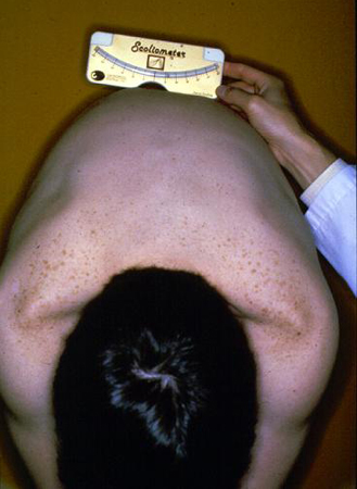
A scoliometer is used to quantify right- and left-sided asymmetries (paraspinal prominences) identified on Adams forward bend test. A positive result is one of >5° at any paraspinal prominence (thoracic or lumbar).
In patients with a flexible spinal deformity, side bending toward the observed paraspinal prominence in the forward flexed position results in decreased magnitude of the observed prominence.[Figure caption and citation for the preceding image starts]: Scoliometer measurement of a right thoracic prominenceWeinstein SL, et al. Adolescent idiopathic scoliosis. Lancet. 2008;371:1527-1537. Used with permission [Citation ends].
Neurologic examination
Involves examination of all 4 extremities.
Includes graded strength testing; assessment of dermatomal sensation, and vibratory and position sense (proprioception); and evaluation of deep tendon reflexes, clonus, and the Babinski reflex.
The gag reflex and supine straight leg raise should also be tested. The presence of a positive supine straight leg raise is evidence of nerve root impingement and is suspicious of an anatomic lesion, which may be contributing to the observed spinal deformity. An abnormal gag reflex may represent an abnormality involving the hindbrain, such as in Arnold-Chiari malformation.
Abdominal reflexes are assessed to rule out intramedullary lesions of upper motor neurons. The umbilicus should deviate toward the abdominal quadrant being stimulated during the examination. Asymmetry or absence of this reflex is suggestive of underlying neuropathology such as syringomyelia.
Assessment of pubertal status
Pubertal status (menarche in girls, development of axillary and facial hair in boys) should also be assessed in order to estimate the time of onset of the peak adolescent growth spurt, as these features occur 6 to 12 months after this period.
Investigations
Plain film radiographs
It is not routinely recommended to obtain x-rays before consulting a spine specialist, as these will routinely be obtained upon the patient's presentation to the specialist.
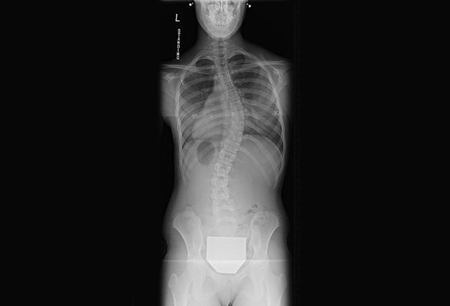
Patients with scoliometer values of 5° or greater (correlates with Cobb angle measurements of at least 10°) warrant further investigation with plain x-rays.[34][35] These should include standing PA and lateral views of the entire spine from the cervicothoracic junction to the hips, including the pelvis, on a single film. [Figure caption and citation for the preceding image starts]: Posteroanterior scoliosis radiograph of a 13-year-old girl with a 49° right thoracic curvature with apex at the T9-T10 disc spaceFrom the collection of Stuart Weinstein, MD, University of Iowa; used with permission [Citation ends].
 [Figure caption and citation for the preceding image starts]: Lateral scoliosis radiograph of a 13-year-old girl with a 49° right thoracic curvatureFrom the collection of Stuart Weinstein, MD, University of Iowa; used with permission [Citation ends].
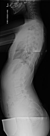
Allow evaluation of the iliac apophysis (Risser sign) and triradiate cartilage in order to calculate bone age and estimate remaining growth potential.[23][24][25][27][36] The iliac apophysis develops from lateral to medial on a coronal view, with the most immature patients having no evidence of an apophysis and the most mature having a fully developed and fused apophysis that extends to the sacroiliac joint. The risk for curve progression in patients with a Risser sign of 1 or less can be as high as 70%.[37] In a patient with a Risser sign of 3, the risk of progression is approximately 10%.[24][25]
On both PA and lateral projections, each cervical/thoracic/lumbar vertebra should be counted, as well as each rib and pedicle bilaterally, in order to identify failures of segmentation or formation that may be the cause of the deformity.
On PA x-rays, Cobb angle measurements should be made for all major and minor curvatures. Cobb angles are measured by determining the most tilted vertebrae at the cranial and caudal end of each curve.[38] These vertebrae are referred to as the end vertebrae (EV). A line is drawn along the upper vertebral end plate of the EV at the top of the curve and along the lower vertebral end plate of the EV at the bottom of the curve. A perpendicular line is then created from each of these lines and the angle formed by the intersection of these perpendicular lines represents the Cobb angle for that particular curvature.
A Cobb angle >10° establishes a diagnosis of scoliosis. The major curve is that with the largest Cobb angle and is always structural. All other curves noted on the PA x-ray are known as minor curves. Minor curves may be structural or compensatory.
PA x-rays also allow evaluation of the degree of truncal decompensation (severe, clinically apparent deformity in which the trunk is no longer centered over the pelvis, resulting from coronal imbalance in severe curves) and overall coronal balance. The extent of coronal decompensation is measured by comparing the distance between the C7 plumb line (C7PL) and the central sacral vertical line (CSVL).
On lateral x-rays, the sagittal balance should be determined by measuring the distance from the C7 plumb line to the posterior superior corner of the S1 vertebral body (the C7PL should pass through the posterior superior corner of S1 on a standing lateral projection). It is also possible to measure the degree of thoracic kyphosis and lumbar lordosis from the standing lateral x-ray.
MRI
Do not order MRI for scoliosis in a child until all appropriate clinical and plain radiographic exams have been completed.[39] An MRI scan of the entire spine and brainstem is recommended in cases where intraspinal pathology (e.g., syringomyelia, Arnold-Chiari malformation, intramedullary tumor/mass) cannot be confidently ruled out as the inciting cause of the spinal deformity through the clinical history, exam, or plain x-rays, as well as in patients with an atypical presentation or abnormality on neurologic exam.
Referral to specialist
Referral to an orthopedic surgeon specializing in pediatric spinal deformity is recommended for patients with paraspinal or rib prominences on the Adams forward bend test and scoliometer measurements of >5°.[34][35]
Use of this content is subject to our disclaimer