Tests
1st tests to order
CT head
Test
Typically normal after mild traumatic brain injury (TBI) but the absence of CT abnormalities does not exclude structural damage.[2] The New Orleans Criteria or the Canadian CT head rule can be used as an assessment guide to imaging.[67][73][74]
Do not routinely obtain CT scans for patients with minor head injury who are at low risk based on validated decision rules.[67][77] In children, use clinical observation/Pediatric Emergency Care Applied Research Network (PECARN) criteria to determine whether imaging is indicated.[78][79]
Several guidelines recommend, or suggest consideration of, CT head imaging for anticoagulated patients after minor head injury, regardless of symptoms.[67][80]
CT is most commonly used because it rapidly rules out intracranial bleeding or bony injuries. The most common structural changes observed are cerebral contusions, and less commonly epidural hematomas, and subdural hematomas.[Figure caption and citation for the preceding image starts]: Sites of contusions and epidural hematomas: (A) prefrontal cortex, (B) pterion, (C) temporal-parietal cortex (often during contre-coup)From the collection of L. Henry; used with permission [Citation ends].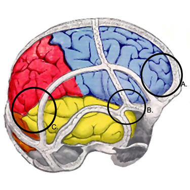 Early detection of epidural and subdural hematomas with CT scanning can be lifesaving.
Result
normal
Tests to consider
MRI head
Test
More likely than CT scan to be abnormal after mild TBI.
Avoid ordering a brain MRI to evaluate an acute concussion unless more severe brain injury is suspected.[82] Brain MRI should be obtained whenever there is suspicion of an intracerebral structural lesion or hematoma, and CT scan is negative. Studies report structural traumatic abnormalities on MRI performed at two-three weeks after injury in approximately 30% of patients with mild TBI who had a normal CT on presentation.[2][26][27] Advanced MRI techniques, such as diffusion tensor imaging and susceptibility weighted imaging are more sensitive than standard MRI at detecting superficial contusions, traumatic axonal injury and traumatic vascular injury.[2][83][84]
Fast MRI is feasible and accurate relative to CT in clinically stable children, where exposure to ionizing radiation may otherwise be a concern.[85]
Result
frequently normal; may show cerebral contusions, epidural or subdural hematomas, traumatic axonal injury and traumatic vascular injury.
Emerging tests
PET, single-photon emission CT (SPECT) of head
multimodal MRI technologies
Test
Diffusion tensor imaging, resting state functional MRI, quantitative susceptibility imaging, magnetic resonance spectrography, and arterial spin labeling are being studied in research protocols aimed at understanding the neurobiologic effects of, and recovery after, mild TBI.[3]
Result
anatomic and metabolic changes in the concussed brain during the acute post-injury phase and through recovery
Use of this content is subject to our disclaimer