Etiology
Etiology is any blow to the head such that the diagnostic criteria for mild traumatic brain injury (TBI) are met.[1][5][6] The injury to the head results in biomechanical forces contorting the brain.[Figure caption and citation for the preceding image starts]: Contortion of the brain during a concussion, based on biomechanical modelingFrom the collection of L. Henry; used with permission [Citation ends].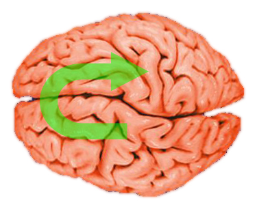 Brain rotation occurs because the head is attached to the neck, on which it pivots when struck or during rapid deceleration. Movement of the brain within the skull is constrained by the falx and anchor points along the skull base and as such, selected areas are subject to high strain forces.[23] These areas, which include the corpus callosum, internal capsule, and fornix, are most likely to undergo axonal injury after mild TBI.[24]
The most common causes of mild TBI include:[25]
Motor vehicle accidents
Being struck by or against objects (includes sports)
Assaults
Falls.
Frequency of cause varies across lifespan.
Pathophysiology
Studies indicate that mild traumatic brain injury (TBI), including concussions, can result in structural changes detectable on CT or structural MRI in up to 30% of all cases.[26][27] The most common structural changes observed are cerebral contusions, and less commonly epidural hematomas, subdural hematomas, and axonal injury.[Figure caption and citation for the preceding image starts]: Sites of contusions and epidural hematomas: (A) prefrontal cortex, (B) pterion, (C) temporal-parietal cortex (often during contre-coup)From the collection of L. Henry; used with permission [Citation ends].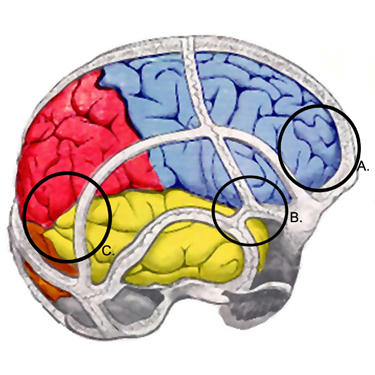 Techniques such as MRI volumetry suggest that mild TBI may lead to global brain atrophy.[28][29]
The biochemical cascade is marked by an initial period of indiscriminate neurotransmitter release and unchecked ionic fluxes.[30] Excitatory neurotransmitters, such as glutamate, bind to N-methyl-D-aspartate receptors leading to further neuronal depolarization with the influx of calcium and the efflux of potassium. These ionic shifts lead to acute and subacute changes in cellular metabolism and physiology. Acutely, the sodium-potassium pump works overtime to restore a homeostatic balance, requiring increasing amounts of adenosine triphosphate and a corresponding increase in glucose metabolism. This hypermetabolic state co-occurs with diminished cerebral blood flow. The disparity in energy balance and the tendency to restore ionic balance is met with decrease in blood flow, creating a cellular energy crisis that is suspected to be the mechanism for post-injury vulnerability, leaving the brain more vulnerable to a second injury and potentially longer-lasting, more severe deficits.
After the initial period of hypermetabolism, the brain goes into a state of hypometabolism where the persistent increase of calcium may be responsible for impairing mitochondria, further affecting cellular metabolism and neural integrity, worsening the energy crisis, and potentially affecting post-traumatic neural connectivity.[30]
Axonal compression and stretching is also thought to be a major injury mechanism, creating a focal abnormality on the surface membrane of the axon within hours of injury, sufficient to impair axoplasmic transport, resulting in edema. [Figure caption and citation for the preceding image starts]: Model of axonal injury after a concussive blowFrom the collection of L. Henry; used with permission [Citation ends].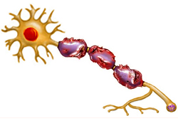 The swollen axon then separates, with the proximal section remaining attached to the cell body while the distal end undergoes phagocytosis by neighboring glial cells. Axonal swellings may persist unchanged, or regeneration may occur over several weeks. If axons do not regenerate, reactive deafferentation may occur.[31]
Neuroimaging studies using functional MRI suggest that a depressed mood after mild TBI may reflect an underlying pathophysiologic abnormality consistent with a limbic-frontal model of depression.[31][32]
Classification
2022 Concussion in Sport Group consensus statement[5]
Several common features may be utilized in clinically defining the nature of a sport-related concussive head injury:
A traumatic brain injury (TBI) caused by a direct blow to the head, neck or body resulting in an impulsive force being transmitted to the brain that occurs in sports and exercise-related activities.
A range of clinical symptoms and signs that may or may not involve loss of consciousness.
Neuropathologic changes, 'but the acute clinical signs and symptoms largely reflect a functional disturbance rather than a structural injury and, as such, no abnormality is seen on standard structural neuroimaging studies.'
Symptoms and signs which may present immediately, or evolve over minutes or hours, and commonly resolve within days, but may be prolonged.
No abnormality seen on standard structural neuroimaging studies (CT or MRI T1- and T2-weighted images); in the research setting, abnormalities may be present on functional, blood flow or metabolic imaging studies.
Injury severity classification of mild traumatic brain injury (MTBI)[6][7]
Criteria used to classify MTBI include:
Structural imaging: normal
Loss of consciousness <30 minutes
Post-traumatic amnesia: 0 to 1 day
Glasgow Coma Scale (GCS): 13 to 15
Abbreviated Injury Scale score (head): 1 to 2.
World Health Organization diagnostic criteria[1]
Operational criteria for MTBI and concussion include:
One or more of confusion or disorientation, loss of consciousness for up to 30 minutes, post-traumatic amnesia for <24 hours, and other transient neurologic abnormalities, including seizure, focal signs, intracranial lesion not requiring surgery
GCS score of 13 to 15
Manifestations must not be due to a penetrating head injury, other injuries (facial, systemic), other problems (psychological trauma, coexisting medical condition), or drugs/alcohol/medication.
Use of this content is subject to our disclaimer