Approach
Adenocarcinoma of unknown primary site (AUP) is characterized by metastatic disease without an identified primary malignancy. If a primary site is found during the diagnostic workup, the focus shifts to treating that specific entity.
A key goal of the diagnostic workup is to determine if a patient falls into one of the more favorable clinicopathologic subtypes of AUP, which would prompt specific treatments.
With appropriate diagnostic tools, 20% to 30% of AUP patients are identified with favorable subtypes, while the majority (70% to 80%) are diagnosed with unfavorable subtypes.
Complete medical history
History of the presenting complaint rarely elucidates any information regarding potential primary sites of disease, as it generally refers to the metastatic foci.
Presenting symptoms and signs commonly include:
Pain from peritoneal or pleural irritation, or pathologic fracture with bony involvement
Localized swelling, if superficial lymph nodes are involved (e.g., cervical chain adenopathy)
Obstructive jaundice due to pancreaticobiliary lesions
Symptoms of postobstructive pneumonia (pneumonia occurring distal to a bronchial mass) (e.g., cough, wheeze, dyspnea)
Hemoptysis with parenchymal lung involvement
Constitutional symptoms (e.g., weakness, anorexia, malaise, early satiety, and weight loss)
Shortness of breath
Neurologic pain or weakness
Headaches and/or seizures
Ascites
Depression
Delirium
Important questions include any history of previous surgery or biopsy, and any history of removed or regressed lesions, and review of existing imaging studies.[15]
A detailed family history should assess the possibility of an inherited cancer syndrome. Smoking and alcohol history and a systems review will help to determine the overall health, or performance status, of the patient.
Physical exam
A thorough physical exam is mandatory, and specific findings may guide further diagnostic workup to seek a likely primary site.
Physical exam should include careful assessment of the thyroid and of lymph node groups, breast exam, and prostate exam in men, or full pelvic exam, with cervical smear as appropriate, in women. Hepatomegaly or a palpable mass may be noted.
Patients presenting with a neck mass should undergo a dedicated head and neck exam.
Laboratory tests
Baseline blood and biochemistry tests are recommended (e.g., complete blood count, electrolytes, liver function, creatinine, calcium).
Fecal occult blood test, lactate dehydrogenase, and urinalysis may be indicated.[15][16]
Biopsy and pathologic diagnosis
The key diagnostic study is a thorough pathologic analysis of a tumor specimen, generally taken from the most easily accessible metastatic focus.
Consult with the pathologist prior to any biopsy to determine the extent of specimen required to differentiate between likely primary sites. Once the specimen is obtained, the morphology of the tumor cells is identified using light microscopy analysis after hematoxylin and eosin staining.
When adenocarcinoma is confirmed, immunohistochemical (IHC) markers are employed to determine likely tumor lineage. This technique involves the application of labeled antibodies to detect specific tumor antigens.[17] A battery of markers is generally applied, although information from the history, physical exam, and initial testing will usually allow a more directed search.[15] Specific tests should be ordered based on indicators from the initial diagnostic workup and in consultation with a pathologist.
No IHC marker is 100% specific, and the information garnered from these tests must be considered in conjunction with the clinical scenario and other test results. However, patterns of IHC staining can be suggestive of likely primary sites in AUP.[15][18][19]
Pan-keratin (AE1/AE3 and CAM5.2): can indicate a carcinoma lineage.
Cytokeratin-7 (CK7): expression is generally limited to lung, breast, ovary, endometrium, and upper gastrointestinal/pancreaticobiliary cancers, and is not seen in lower gastrointestinal tract tumors.
Cytokeratin-20 (CK20): generally expressed on gastrointestinal epithelium, urothelium, and Merkel cells.
CDX2 or SATB2: suggest colorectal or other gastrointestinal primary.
TTF-1: suggestive of lung or thyroid primary.
Napsin A: suggests lung primary.
GATA3: suggests breast/urinary bladder/salivary gland primary.
A more extensive list of IHC markers is available in published guidelines.[15][19]
If initial IHC markers narrow the differential of likely primary sites, more specific antibodies can be utilized to further solidify the diagnosis.[20] Results may not, however, alter therapeutic recommendations; caution must be taken to avoid unnecessary delay to treatment initiation.
IHC marker profiles may occasionally differ between primary and metastatic lesions.[21][22][23]
Identification of primary site
Targeted treatment can lead to improved outcomes.
In women, workup should evaluate for an occult primary breast or ovarian tumor.
IHC markers, hormone receptor status, serum tumor markers, and dedicated imaging, including mammography, breast magnetic resonance imaging (MRI), and transvaginal ultrasound, should be undertaken.
Women with isolated axillary adenopathy should undergo extensive workup for occult breast carcinoma, including mammography ± breast MRI/ultrasound, and estrogen/progesterone (ER/PR) hormone receptor status on tumor cells.
Paracentesis (if technically possible) is recommended in all patients presenting with ascites.[24] Female patients with ascites histology consistent with ovarian cancer are responsive to tumor debulking and chemotherapy.[2][25][26][27]
In men, workup should focus on identification of primary prostate or germ cell tumor.
Further investigations
If a likely primary site is suggested by the history and physical exam, diagnostic testing should focus on this. However, in the vast majority of cases, testing will not be so directed. In general, further clinical investigations will comprise pathology, imaging, and endoscopy studies; caution must be taken to avoid unnecessary delay to treatment initiation. Since the diagnostic yield is often low, the benefit to the outcome of the patient may be marginal.[30]
Radiographic studies
CT chest/abdomen/pelvis with intravenous contrast (unless contraindicated) for initial staging is recommended.[3][15][Figure caption and citation for the preceding image starts]: CT abdomen with intravenous contrast, revealing numerous enhancing lesions in the right hepatic lobe, with associated ascites. Percutaneous biopsy of one of these hepatic lesions revealed adenocarcinoma, but no primary site was identified during routine workup. This is a typical presentation of AUPFrom the personal collection of Dr D. Cosgrove [Citation ends].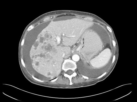 [Figure caption and citation for the preceding image starts]: CT abdomen with intravenous contrast, revealing numerous enhancing liver lesions in both hepatic lobes. Percutaneous biopsy of a right lobe lesion revealed adenocarcinomaFrom the personal collection of Dr D. Cosgrove [Citation ends].
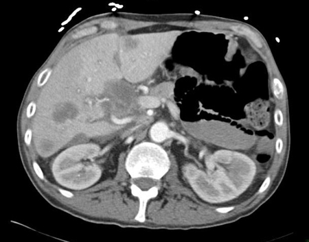
Mammography is required for patients with clinical features compatible with breast cancer.[15] Breast MRI is indicated (without or with contrast [unless contraindicated]) if mammography is not diagnostic and/or in women with adenocarcinoma identified in axillary lymph nodes.[3][15]
Patients presenting with a neck mass undergo fiberoptic exam, computed tomography (CT), and 18F-fluorodeoxyglucose-positron emission tomography/CT (FDG-PET/CT) as clinically indicated.[16] FDG-PET/CT is not routinely recommended in evaluation of cancers of unknown primary; it may be considered in those for whom contrast enhancement is contraindicated.[3][15]
Transvaginal ultrasound is the preferred imaging modality for women presenting with ascites to assess for the presence of an ovarian mass. Color or power Doppler should be included in the exam.[31]
Endoscopic studies
Gastroesophageal or colorectal endoscopic exam is not uniformly necessary in asymptomatic patients with AUP. However, endoscopy should be considered in patients with IHC tests suggestive of an upper gastrointestinal or colorectal primary, or in selected patients premised upon clinical suspicion.
Direct laryngoscopy with or without esophagoscopy and bronchoscopy is recommended for patients with cervical lymphadenopathy. The extent of exam is dependent on the level of cervical nodes involved. Additional IHC testing can help to confirm an occult head and neck primary tumor.
Serum tumor markers
Assessment of serum tumor markers may be considered in specific clinical scenarios. However, they are nonspecific and have no diagnostic role in patients with AUP.
Prostate-specific antigen (PSA): men over the age of 40 years with AUP, and all men with AUP and bone metastases, should have PSA checked to assess for potential prostate carcinoma.
Serum alpha-fetoprotein (AFP)/beta-human chorionic gonadotropin (beta-hCG): may be indicative of germ cell tumors, and should be checked in patients with mediastinal tumors and in men under the age of 65 years presenting with a retroperitoneal mass.
CA 125: typically elevated in women with histology consistent with ovarian cancer.
Estrogen and progesterone receptor status
IHC markers for estrogen receptor (ER) and progesterone receptor (PR) should be performed in all women with adenocarcinoma, especially in those with isolated axillary lymphadenopathy.
Assessments of carcinoembryonic antigen (CEA), CA 125, CA 19-9, and CA 15-3 should not be ordered as part of a nondirected diagnostic workup. They may, however, have a role in assessing treatment response in individual patients; consideration may be given to establishing a baseline at initial testing.
Next-generation sequencing (NGS)
Can identify potentially actionable mutations and aberrations (e.g., NTRK or RET gene fusions), and should be considered after initial determination of histology is completed.[15]
Testing for predictive biomarkers (including tumor mutational burden [TMB], and assessment of microsatellite instability resulting from deficient DNA mismatch repair [MSI/dMMR]) may help to identify potentially actionable mutations.[15] One randomized study of targeted therapy (guided by comprehensive genomic profiling, an NGS approach) reported improved progression free survival in patients with unfavorable cancer of unknown primary who were treated with molecularly guided therapy rather than standard chemotherapy (6.01 months vs. 4.4 months, respectively).[32] Overall survival data were immature at cutoff.
Emerging investigations: gene expression profiling
Gene expression profiling (GEP) encompasses molecular assays that employ messenger RNA-, DNA-, or microRNA-based platforms to identify cancers of unknown primary. The technology is premised on the assumption that the molecular profile of the metastatic tumor(s) has a similar profile to that of the primary tumor. Surrogate measures (e.g., IHC results) are typically used to determine assay accuracy because the primary site of AUP is typically difficult to determine.
Observational studies suggest clinical utility.[33][34] However, prospective randomized clinical trials and meta-analyses of (prospective and retrospective) studies investigating the role of molecularly defined site-specific treatment of cancers of unknown primary have failed to report significant improvement in overall survival compared with empiric therapy.[35][36]
The clinical benefits of GEP testing remain to be determined; guidelines do not currently recommend routine GEP testing in patients with AUP.[2][15]
Use of this content is subject to our disclaimer