Images and videos
Images

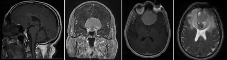
Meningioma
Sagittal image (left) demonstrates large extra-axial mass isointense with brain. After contrast administration, the lesion avidly enhances, as shown in the coronal image (center left) and axial image (center right). Note the extensive edema surrounding the tumor on the T2 axial image (right)
From the personal library of Dr William T. Couldwell; used with permission
See this image in context in the following section/s:

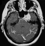
Meningioma
Axial contrast-enhanced image demonstrates meningioma in the cavernous sinus on the left side
From the personal library of Dr William T. Couldwell; used with permission
See this image in context in the following section/s:
Use of this content is subject to our disclaimer