Tests
1st tests to order
Dix-Hallpike maneuver
Test
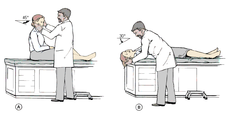
Also referred to as the Nylen-Barany maneuver. Used to diagnose posterior canal BPPV. The patient is seated and positioned on an examination table such that the patient's shoulders will come to rest on the top edge of the table when supine, with the head and neck extending over the edge.
The patient's head is turned 45° toward the ear being tested.
The head is supported, and then the patient is quickly lowered into the supine position with the head extending about 30° below the horizontal while remaining turned 45° toward the ear being tested.
The head is held in this position and the physician checks for nystagmus.
To complete the maneuver, the patient is returned to a seated position and the eyes are again observed for reversal nystagmus.
The Dix-Hallpike maneuver can also be used to diagnose the rare anterior canal variant of BPPV, but the fast phase of nystagmus would be down-beating, opposite to posterior canal BPPV of the same ear; the torsional component would be in a similar direction, although more subtle. A straight head hanging position has also been described to diagnose anterior canal BPPV, and it involves moving the patient from a sitting to lying position, with the head tilted (extended) straight back. The torsional component would suggest whether the right or left anterior canal is affected, similar to that of posterior canal BPPV of the same ear.[38][Figure caption and citation for the preceding image starts]: Dix-Hallpike maneuverParnes LS, Agrawal SK, Atlas J. Diagnosis and management of benign paroxysmal positional vertigo (BPPV). CMAJ. 2003:169:681-693. Used with permission [Citation ends].
Result
vertigo with the appropriate position-provoked nystagmus response; the nystagmus and vertigo occur with 1 to 5 seconds of latency and last <30 seconds; nystagmus is torsional (rotatory) in nature, reversible with sitting, and fatigable with repeat testing; left ear BPPV has a clockwise torsional nystagmus response, while right ear BPPV has a counterclockwise response
supine lateral head turns
Test
Used to diagnose lateral (horizontal) canal BPPV.
The clinician places the patient in a supine position and, ideally, flexes the neck 30° from horizontal to bring the lateral canals into the vertical plane of gravity. However, it is sufficient and more usual to simply lay the patient flat on their back.
The head is then rotated to one side, left for 1-2 minutes, and then rotated to the opposite side.
Similar to the Dix-Hallpike maneuver, a positive test is noted when the patient experiences vertigo with nystagmus.
Depending on the pathophysiologic process, different responses may be observed.[Figure caption and citation for the preceding image starts]: Lateral (horizontal) canal BPPVParnes LS, Agrawal SK, Atlas J. Diagnosis and management of benign paroxysmal positional vertigo (BPPV). CMAJ. 2003:169:681-693. Used with permission [Citation ends].
In general, the vertigo and nystagmus of lateral canal BPPV has a shorter latency, is less fatigable on repeat testing, can be more severe, and is more often associated with vomiting, as compared with posterior.[7][37]
Result
horizontal nystagmus without a torsional (rotatory) component; apogeotropic nystagmus (away from the ground) indicates cupulolithiasis, and geotropic (toward the ground) nystagmus indicates canalithiasis; the side with the stronger response corresponds to the affected canal in canalithiasis, and the weaker response corresponds to the affected canal in cupulolithiasis
Tests to consider
audiogram
Test
Indicated in patients with hearing loss. Primary BPPV is not associated with hearing loss, but an audiogram can be helpful in diagnosing other pathophysiologic processes, secondary BPPV, or a coexisting disorder.
Result
normal in primary BPPV, unless coexisting condition present; can be abnormal in secondary BPPV; in patients with Meniere disease, an audiogram will demonstrate a sensorineural hearing loss, usually unilateral and initially worse in the low frequencies; labyrinthitis will also demonstrate sensorineural hearing loss on an audiogram
brain MRI
Test
MRI and MRI angiography can be useful in diagnosing or excluding central nervous system conditions such as multiple sclerosis, posterior fossa tumors, and ischemic processes that may mimic BPPV.
Result
findings indicative of a central condition that may mimic BPPV
Use of this content is subject to our disclaimer