Tests
1st tests to order
radiograph of a long bone
Test
Plain film of knees and wrists are taken when rickets is suspected.[4][Figure caption and citation for the preceding image starts]: Right wrist of a patient with vitamin D deficient rickets before treatment. His right wrist x-ray showed sclerotic and widened end plates of the radius and ulna (arrows)Seerat I, Greenberg M. Hypocalcaemic fit in an adolescent boy with undiagnosed rickets. BMJ Case Reports 2010; doi:10.1136/bcr.10.1136/bcr10.2008.1153 [Citation ends].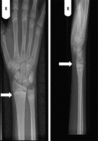
Result
widening of the epiphyseal plate, loss of definition of the zone of provisional calcification at the epiphyseal/metaphyseal interface, cupping, splaying, and fraying of the metaphysis; Looser zone (pseudofracture)
serum calcium
serum inorganic phosphorus
serum parathyroid hormone level
25-hydroxyvitamin D levels (calcidiol)
Test
Calcitriol (1,25-dihydroxyvitamin D) is the active form of vitamin D, but calcidiol (25-hydroxyvitamin D) is preferred for testing as it has a longer half-life and is found at much higher levels in the serum.
Normal values >10.0 nanograms/mL (>25 nanomoles/L).[3]
Result
low in vitamin D-deficient rickets, usually <10 nanograms/mL (<25 nanomoles/L); normal in genetic forms of hypocalcemic rickets and in hypophosphatemic rickets
alkaline phosphatase and liver function tests
serum creatinine and blood urea nitrogen
Test
Kidney disease causes abnormal calcium and phosphorus regulation and impaired synthesis of calcitriol.[4]
Result
elevated in rickets caused by kidney disease
urinary calcium and phosphorus
Test
Serum and urine values are used to calculate percent tubular reabsorption of phosphate (TRP). Low TRP is diagnostic of hypophosphatemic rickets in the absence of vitamin D deficiency.[25] Tubular reabsorption of phosphate is normally >80%.[24]
Result
urinary calcium is decreased and urinary phosphorus is increased in hypocalcemic rickets; urinary calcium is normal and urinary phosphorus is high in hypophosphatemic rickets
Tests to consider
1,25-dihydroxyvitamin D levels (calcitriol)
Test
Normal values 16.5 to 53.5 picograms/mL (43-139 picomoles/L); up to 96.2 picograms/mL (250 picomoles/L) in preterm babies.[3][5]
Calcitriol may be normal, low, or high in relation to the reference range. Serum calcitriol concentration is inappropriately low for the prevailing phosphate level in patients with X-linked hypophosphatemic rickets (hypophosphatemia typically stimulates calcitriol synthesis), and very low in type I vitamin D-dependent rickets (pseudovitamin D-deficient rickets). In patients with type II vitamin D-dependent rickets (end-organ resistance to calcitriol), serum calcitriol concentration is usually very high.
Result
typically normal or elevated in hypocalcemic rickets as a result of parathyroid hormone action; usually normal in hypophosphatemic forms of rickets
Use of this content is subject to our disclaimer