History and exam
Key diagnostic factors
common
pruritus
Intense pruritus is present in 80% of patients with cutaneous lesions.[53]
violaceous, flat-topped papules or plaques
Typically present in patients with cutaneous lesions.
Lesions are most commonly found on the flexor wrists and ankles but also on the trunk and extremities. May occur on the vulva in genital disease.[Figure caption and citation for the preceding image starts]: Cutaneous lichen planusMayo Clinic clinical photographs (used with permission) [Citation ends].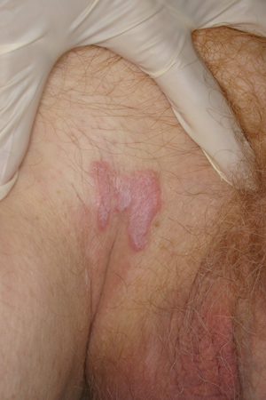
Wickham striae
Overlying lacy white networks may be seen in cutaneous lesions.
mucosal erosions and lacy white network
Present in 18% of patients with oral or genital lesions.[26]
Classically, white reticulation of buccal mucosa, painful desquamative gingivitis, and tongue erosions are seen in oral disease.[3]
Characteristic features of erosive vulvar LP include a well-demarcated, glazed red macule or patch, which may be edged by a white lacy or raised border, located on the labia minora, vestibule, and/or vagina.[50][Figure caption and citation for the preceding image starts]: Oral lichen planusMayo Clinic clinical photographs (used with permission) [Citation ends].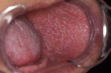 [Figure caption and citation for the preceding image starts]: Genital lichen planusMayo Clinic clinical photographs (used with permission) [Citation ends].
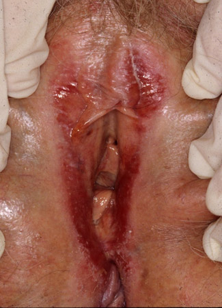
uncommon
scarring alopecia
Typically seen in patients with lichen planopilaris (LP of the scalp).
Follicular keratotic plugs with perifollicular inflammation are seen at onset. Scarring alopecia ensues.[5][6][7][Figure caption and citation for the preceding image starts]: Lichen planopilarisMayo Clinic clinical photographs (used with permission). [Citation ends].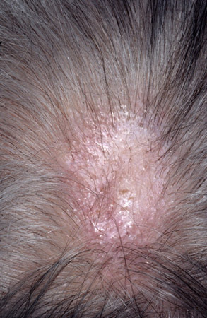
nail involvement
Nail deformity, including lateral thinning of the nail plate, longitudinal ridging, dorsal pterygium, and scarring is evident in 15% of patients.[26][Figure caption and citation for the preceding image starts]: Ungual lichen planusMayo Clinic clinical photographs (used with permission) [Citation ends].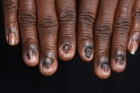 [Figure caption and citation for the preceding image starts]: Ungual lichen planusMayo Clinic clinical photographs (used with permission) [Citation ends].
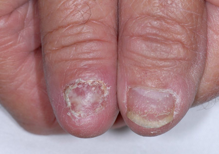
Other diagnostic factors
uncommon
dorsal pterygium
Cuticle growth over the nail may be evident.
history of hepatitis C (HCV)
Hepatitis C may induce susceptibility by changing cytokine expression in some patient populations.[30]
Almost 20% of newly diagnosed patients may be hepatitis C positive, according to one study.[30] One systematic review reported a global prevalence of hepatitis C among patients with LP of 9.42%.[42]
Risk factors
strong
hepatitis C (in some patient populations)
Hepatitis C may induce susceptibility by changing cytokine expression in some patient populations.[30]
There is an established association between hepatitis C and lichen planus.[42] One study shows almost 20% of newly diagnosed LP patients are hepatitis C positive.[30]
One systematic review reported a global prevalence of hepatitis C among patients with LP of 9.42%.[42]
weak
-308 G/A polymorphism in TNF-alpha gene
The -308 G/A polymorphism in the TNF-alpha gene may be a genetic risk factor for oral LP in patients without hepatitis C infection and those of mixed ethnicity.[43]
hepatitis B vaccination
There are case reports of disease arising after hepatitis B vaccination.[17]
influenza vaccination
There are case reports of disease arising after inactivated influenza vaccination.[16]
coronavirus disease 2019 (COVID-19) vaccination
There are case reports of disease arising after mRNA COVID-19 vaccinations.[18]
psychosocial stress
It has been demonstrated that the onset and extension of lesions maybe induced by stressful situations, especially those involving family.[44]
oral allergens
Oral allergens (including metals, plastics, and flavorings) are well recognized causes of oral disease and have been demonstrated.[31]
use of certain drugs
Consider correlations between lesion onset and initiation of any LP-associated drugs; the most frequently associated drugs include ACE inhibitors, thiazide diuretics, antimalarials, beta-blockers, gold salts, penicillamine, immune checkpoint inhibitors, tyrosine kinase inhibitors, and tumor necrosis factor (TNF)-alpha antagonists. Other drugs have been associated with lichenoid eruptions of the oral mucosa (e.g., allopurinol, ketoconazole, nonsteroidal anti-inflammatory drugs [NSAIDs], anticonvulsants, antiretrovirals).[34][45]
Use of this content is subject to our disclaimer