Images and videos
Images
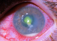


Keratitis
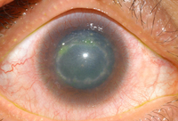
A ring ulcer due to Acanthamoeba after start of treatment
Courtesy of F. I. Proctor Foundation, UCSF
See this image in context in the following section/s:

Keratitis
Moraxella corneal ulcer
Courtesy of F. I. Proctor Foundation, UCSF
See this image in context in the following section/s:
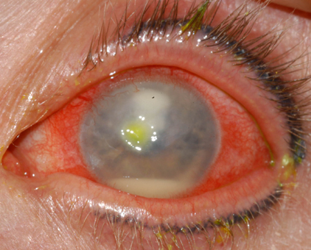
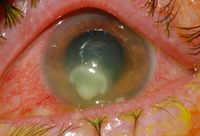
Keratitis
Fusarium keratitis with hypopyon
Courtesy of F. I. Proctor Foundation, UCSF
See this image in context in the following section/s:

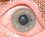
Keratitis
Early, superficial Acanthamoeba keratitis
Courtesy of F. I. Proctor Foundation, UCSF
See this image in context in the following section/s:

Keratitis
Fusarium corneal ulcer showing typical feathery edges and a satellite lesion
Courtesy of F. I. Proctor Foundation, UCSF
See this image in context in the following section/s:

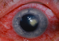
Keratitis
Bacterial corneal ulcer with hypopyon
Courtesy of F. I. Proctor Foundation, UCSF
See this image in context in the following section/s:
Use of this content is subject to our disclaimer