Images and videos
Images


Renal artery stenosis
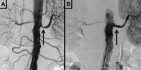
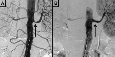
Digital subtraction angiography in a patient with significant atherosclerotic left renal artery stenosis. Panel A, prior to stent placement. Panel B, after successful stent deployment. Arrows indicate the site of stenosis and stent placement in their respective panels
Courtesy of Alvaro Alonso, MD and Scott J. Gilbert, MD
See this image in context in the following section/s:
Renal artery stenosis
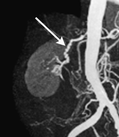
Magnetic resonance angiography (maximum-intensity projection) in a patient with fibromuscular dysplasia of the renal arteries. Arrow indicates the characteristic irregular contour in the right renal artery
Courtesy of Raul Galvez, MD, MPH and Hale Ersoy, MD; Department of Radiology, Brigham and Women’s Hospital, Harvard Medical School
See this image in context in the following section/s:
Renal artery stenosis
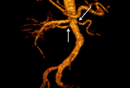
Magnetic resonance angiography (3-dimensional volume rendered reconstruction) in a patient with significant bilateral atherosclerotic renal artery stenosis. Arrows indicate proximal bilateral stenoses
Courtesy of David J. Sheehan, DO; Radiology Department, University of Massachusetts Medical Center and Medical School
See this image in context in the following section/s:
Use of this content is subject to our disclaimer