Urgent considerations
See Differentials for more details
Acutely impaired or deteriorating level of consciousness constitutes an emergency. Such patients need to be assessed urgently from both neurological and general (especially cardiovascular-pulmonary) perspectives. It is usually necessary to have a team of doctors and nurses tackle these problems simultaneously. Airway protection and attention to vital sign abnormalities take priority.
Airways management
In the unconscious patient, the airway may be blocked due to obstruction of the pharynx by the tongue, vomit, or a foreign body. The airway should be cleared by suction and by “head tilt and chin lift” technique or the modified “chin thrust” if there is concern about cervical spine stability. Lying the patient on his or her side is often sufficient for the patient obtunded after a convulsive seizure, as respirations should be adequate and consciousness often recovers quickly without the need for an oral airway (may induce vomiting) or endotracheal intubation. If it is anticipated that the patient may remain comatose for a longer time, or if there already has been aspiration of secretions, an endotracheal tube is placed. Adequate oxygenation often requires the supplemental administration of higher percentages of oxygen (e.g., 60% to 100%).
Shock
The patient with shock will look unwell and often have symptoms specific to the underlying cause (e.g., fever, chest pain, shortness of breath, or abdominal pain). This may be difficult to recognize in practice.
Shock, with impaired tissue perfusion, sometimes precedes a drop in blood pressure. The following signs raise the suspicion of shock and impending blood pressure fall: tachycardia, cool extremities, weak peripheral pulses, prolonged capillary refill (>2 seconds), and narrowing of the pulse pressure (<25 mmHg). Restoring blood volume and providing inotropic support are often necessary. Vasopressors can help raise the blood pressure, but might impair peripheral perfusion.
Overdose
Signs of opioid overdose include small, reactive pupils, hypoventilation, bradycardia, and hypothermia. Such patients need prompt airway, respiratory, and circulatory support, including endotracheal intubation, intravenous volume replacement, and the administration of an opioid antagonist. Naloxone is usually given intravenously in repeated doses. The effect is usually prompt recovery, but it may be short-lasting.
Overdose with tricyclic antidepressants (TCA), such as amitriptyline, in addition to causing impaired consciousness (from blocking the reuptake of various aminergic neurotransmitters), produces prominent anticholinergic effects. Such patients may develop seizures, bradycardia, hypotension, and various ventricular arrhythmias. The ECG can provide clues for TCA toxicity: the last 40 msec of the QRS complex shows a right axis deviation; R wave >S wave amplitude or >3 mm in aVR; and prolongation of the QRS complex. In addition to activated charcoal through the nasogastric tube, respiratory, airway, and circulatory support, and repeated doses of physostigmine, can help reverse the anticholinergic toxicity. Cardiac pacing is sometimes necessary. Seizures may require antiepileptic drug therapy.
Wernicke encephalopathy
There are usually preserved pupillary reflexes with ocular movement palsies of absent vestibular-ocular reflexes.[42][43] This is a classic presentation but can be mimicked by drug overdose. The triad of ataxia, ophthalmoplegia, and encephalopathy is not always present. Wernicke encephalopathy may also feature with hypothermia and coma, and in patients with histories compatible with vitamin B deficiency (alcoholic people, nutritionally deprived patients, those with gastric stapling, or patients on hemodialysis not taking supplemental B vitamins). Blood tests for pyruvate and erythrocyte transketolase are important, and parenteral thiamine (50-100 mg) is required promptly. Carbohydrate loading should be avoided until this is done. MRI shows increased signal on fluid-attenuated inversion recovery (FLAIR) in mammillary bodies, hypothalamus, medial thalamus, and floor of fourth ventricle. Treatment includes stabilization and resuscitation with airway protection if necessary, high-dose parenteral thiamine, correction of magnesium deficiency, and multivitamin supplementation.
Head injury
Evidence of trauma is often available historically, but not always. Bruising should be looked for, especially linear bruising. Signs of significant head trauma, with basal skull fractures, include hemotympanum, Battle sign (bruising over the mastoids), and raccoon eyes (indicating a fracture of the orbital roof).
In the “talk and die” syndrome, the patient has concussion, recovers consciousness (lucid interval), and then deteriorates to stupor and then coma, usually due to acute subdural or epidural hematoma. It is important to look for lateralized findings, such as gaze preference or conjugate eye deviation to one side, asymmetry of limb movements or frank hemiplegia, and/or pupillary asymmetry (usually there is dilation of the pupil ipsilateral to the lesion; note that about 20% of people have anisocoria). This is followed by loss of ipsilateral pupillary reactivity and paralysis of adduction of the eye. In the later stages the opposite pupil loses its reactivity, due to intrinsic midbrain damage from herniation.[44] Prompt evaluation and action including supportive airway and blood pressure care is required. Urgent CT is indicated. Recognizing early phases of herniation syndromes is important, and investigations and treatments should begin before irreversible damage occurs. Treatment with intravenous mannitol or hypertonic saline and other measures (elevation of the head by 30° in a midline position; supportive ventilation) can provide time before neurosurgical intervention and can be life-saving. Current guideline recommendations on the management of people with traumatic brain injury should be followed.[17][18] See Mild traumatic brain injury and Skull fractures.
[Figure caption and citation for the preceding image starts]: Battle’s sign: superficial ecchymosis over the mastoid processvan Dijk GW. Practical Neurology. 2011;11(1):50-55; used with permission [Citation ends].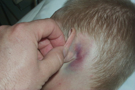
Basilar artery thrombosis
Symptoms are of occipital lobe ischemia (photopsia, visual loss) and signs include quadriparesis, pseudobulbar palsy, pupillary palsies, and ocular palsies.[45] MRI or CT angiogram shows basilar artery occlusion, either in the proximal portion (top of the basilar syndrome, usually with midbrain and thalamic damage) or more extensively. Treatment in early presentations is thrombolysis with intravenous or intra-arterial recombinant thromboplastin activator (rTPA). This can reverse the ischemic damage, which is often fatal if untreated.
Hypoglycemia
Cold perspiration, confusion, or lightheadedness/agitation precedes loss of consciousness. Seizures, multifocal or generalized, should also prompt consideration or screening for hypoglycemia.[46] Point-of-care testing usually allows for recognition of hypoglycemia.
Prompt administration of dextrose is required. In the comatose patient, give intravenous dextrose immediately if the patient has intravenous access.[47][48][49] If intravenous access is not available (e.g., in an outpatient setting), give glucagon or dasiglucagon, although dextrose is preferred if available.[47][49][50]
If the patient has recurrent episodes of hypoglycemia, give a dextrose intravenous infusion in order to sustain euglycemia.[51]
In the patient who may be nutritionally deprived it is wise to pre-administer thiamine to prevent Wernicke encephalopathy.
Myxedema coma
Patients often appear pale and edematous, and there is often puffiness around the eyes. Systemically, they may be hypothermic and have respiratory failure with hypercarpnia. There is often a past history of hypothyroidism, treated hyperthyroidism, head injury, or pituitary surgery (central cause). Serum thyroxine (T4) and triiodothyronine (T3) are depressed. Serum thyroid-stimulating hormone (TSH) is elevated in thyroid gland failure, while TSH is abnormally low in central hypothyroidism. Replacement of T4 or T3 should be incremented gradually in patients with cardiovascular disease. In cases of central hypothyroidism, or if hypothyroidism is longstanding, pretreatment with corticosteroids such as hydrocortisone is recommended to avoid "adrenal crisis."
Cerebral venous thrombosis
It is important to consider this as a diagnosis when patients present with headache of subacute onset that is intractable and worsening, and often associated with nausea and vomiting.[52] Seizures are also common if the cortical vein is involved. Deep cerebral vein involvement leads to coma with thalamic involvement. Urgent MRI (with venous phase) or CT angiogram (with venous follow-through) is required. Prompt recognition and heparinization or thrombolysis is often life-saving. Anticoagulation is required and used even in the presence of intraparenchymal hemorrhage.[53] Continued thrombosis, cerebral edema, and hemorrhage will continue if untreated. Thrombolysis is considered in extreme situations (e.g., extensive thrombosis of deep cerebral venous system). Anticonvulsants are used for seizures.
Bacterial meningitis
This should be considered in the presence of any 2 of: fever, headache, nuchal rigidity, or any alteration in mental status before coma.[54] Children also often have vomiting, photophobia, and lethargy. A petechial rash raises the possibility of meningococcal meningitis. Blood cultures and empirical antibiotics should be initiated while requesting CT or MRI scan. If neuroimaging is negative for mass effect, lumbar puncture is required. Empirical therapy is often indicated initially, until the specific organism and its sensitivities are known. If indicated, empirical therapy should be started immediately according to local sensitivities and protocols. See Bacterial meningitis in adults and Meningococcal disease.
Purulent meningitis is usually fatal if not treated and carries a high morbidity if treatment is delayed. For penicillin-sensitive Streptococcus pneumoniae, penicillin G, cefepime, ceftriaxone, or cefotaxime is suitable. For penicillin-resistant forms the latter 3 are used. For Neisseriameningitidis, penicillin G or ampicillin is suitable. For Haemophilus influenzae, ceftriaxone, cefotaxime, or cefepime is suggested. These should be given intravenously at appropriate doses for 10 to 14 days.
Encephalitis
An acute or subacute onset of a febrile illness, altered mental status, focal neurological abnormalities, and seizures raises suspicion for this condition. This is a medical emergency; hence, management consists of basic resuscitation measures ensuring adequacy of the airway, breathing, and circulation, and empirical antiviral therapy in cases of suspected viral encephalitis concurrently with diagnostic steps. All suspected cases of encephalitis should be admitted and fully evaluated. Some patients with milder symptoms and signs can be managed in a regular nursing unit, with access to an intensive care unit (ICU) bed if needed. All other patients, and in particular those with complications (e.g., significant electrolyte abnormalities, strokes, elevated intracranial pressure, cerebral edema, coma, seizure activity, or status epilepticus) should be managed in an ICU, preferably a neuro-intensive care unit.
Prompt isolation is required for all forms of encephalitis until the etiology is determined; encephalitides with airborne or contact transmission to immunocompetent hosts (herpes simplex virus [HSV], varicella, mumps, rubella, enteroviruses, upper respiratory viral infections) require mandatory isolation. Less common infectious causes should involve close collaboration between the clinicians and microbiologists/virologists.
Etiology is often obscure, and therefore no specific measures exist for the majority of the cases. However, for cases where a diagnosis is reasonably certain, treatment is directed toward the underlying offending agent (e.g., antivirals for viral encephalitis). Even when the diagnosis is certain, treatments are not available for many of the encephalitides. All cases of suspected community-acquired viral encephalitis are started empirically on acyclovir until the infecting virus is determined. In an immunocompromised patient, cytomegalovirus encephalitis is a consideration. If suspected, ganciclovir and foscarnet are given with acyclovir until a viral cause is either confirmed or excluded.
Sepsis
Sepsis is a spectrum of disease, where there is a systemic and dysregulated host response to an infection.[55]
Presentation ranges from subtle, nonspecific symptoms (e.g., feeling unwell with a normal temperature) to severe symptoms with evidence of multiorgan dysfunction and septic shock. Patients may have signs of tachycardia, tachypnea, hypotension, fever or hypothermia, poor capillary refill, mottled or ashen skin, cyanosis, newly altered mental state, or reduced urine output.[56]
Risk factors for sepsis include: age under 1 year, age over 75 years, frailty, impaired immunity (due to illness or drugs), recent surgery or other invasive procedures, any breach of skin integrity (e.g., cuts, burns), intravenous drug misuse, indwelling lines or catheters, and pregnancy or recent pregnancy.[56]
Early recognition of sepsis is essential because early treatment improves outcomes.[56][57][Evidence C][Evidence C] However, detection can be challenging because the clinical presentation of sepsis can be subtle and nonspecific. A low threshold for suspecting sepsis is therefore important. The key to early recognition is the systematic identification of any patient who has signs or symptoms suggestive of infection and is at risk of deterioration due to organ dysfunction.
Screening tools
Several risk stratification approaches have been proposed. All rely on a structured clinical assessment and recording of the patient’s vital signs.[56][58][59][60][61] It is important to check local guidance for information on which approach your institution recommends. The timeline of ensuing investigations and treatment should be guided by this early assessment.[60]
Sepsis screening tools are designed to promote early identification of sepsis and consist of manual methods or automated use of the electronic health record (EHR). These include the Sequential (or Sepsis-related) Organ Failure Assessment (SOFA) score, the quick SOFA (qSOFA) criteria, National Early Warning Score (NEWS), and Modified Early Warning Score (MEWS). There is wide variation in diagnostic accuracy of these tools but they are an important component of identifying sepsis early for timely intervention.[57]
The Third International Consensus Group (Sepsis-3) recommends using the SOFA score (primarily validated in patients in intensive care), with a score ≥2 in a patient with a suspected infection being suggestive of sepsis.[55]
Although the presence of a positive qSOFA should alert the clinician to the possibility of sepsis in all resource settings, its poor sensitivity has led the Surviving Sepsis Campaign to advise against using the qSOFA compared with National Early Warning Score (NEWS), or Modified Early Warning Score (MEWS) as a single screening tool for sepsis or septic shock.[57]
The National Institute for Health and Care Excellence (NICE) UK guideline on sepsis emphasises the need to ‘think sepsis’ in any patient presenting with possible infection. It recommends structured observations and stratification of risk of severe illness and death according to patient age and setting.[56]
Management of patients with suspected sepsis
Treatment guidelines have been produced by the Surviving Sepsis Campaign and remain the most widely accepted standards.[57][62]
Recommended treatment of patients with suspected sepsis is:
Measure lactate level, and remeasure lactate if initial lactate is elevated (>18 mg/dL [>2 mmol/L]).
Obtain blood cultures before administering antibiotics.
Administer broad-spectrum antibiotics (with methicillin-resistant Staphylococcus aureus [MRSA] coverage if there is high risk of MRSA) for adults with possible septic shock or a high likelihood for sepsis.
For adults with sepsis or septic shock at high risk of fungal infection, empiric antifungal therapy should be administered.
Begin rapid administration of crystalloid fluids for hypotension or lactate level ≥36 mg/dL (≥4 mmol/L). Consult local protocols.
Administer vasopressors peripherally if hypotensive during or after fluid resuscitation to maintain MAP ≥65 mm Hg, rather than delaying initiation until central venous access is secured. Norepinephrine (noradrenaline) is the vasopressor of choice.
For adults with sepsis-induced hypoxemic respiratory failure, high flow nasal oxygen should be given.
Ideally these interventions should all begin in the first hour after sepsis recognition.[62]
For adults with possible sepsis without shock, if concern for infection persists, antibiotics should be given within 3 hours from the time when sepsis was first recognized.[57] For adults with a low likelihood of infection and without shock, antibiotics can be deferred while continuing to closely monitor the patient.[57]
See Sepsis in adults and Sepsis in children.
Hypophosphatemia
Hypophosphatemia (refeeding syndrome) most commonly occurs in malnourished patients who are fed in the hospital, causing a shift of phosphate ions from the blood to the intracellular compartment.[63] Marked lowering of serum phosphate, usually to <1.5 mg/dL or 0.5 mmol/L, can produce an acute encephalopathy with seizures, myoclonus, and coma. Severe weakness due to a profound myopathy may necessitate assisted ventilation. It is important to measure serum phosphate in malnourished patients who develop the above features and, if phosphate is low, to replace it intravenously.
Posterior reversible encephalopathy syndrome (PRES)
PRES may be caused by hypertensive encephalopathy, acute hypertension of pregnancy, sepsis, and certain drugs (e.g., cyclosporine and tacrolimus).[64] It occurs with acute/subacute elevations of blood pressure to levels that overcome cerebral autoregulation (e.g., 240/130 mmHg). This may produce vasogenic edema, usually most marked in the white matter of the posterior parts of the cerebral hemispheres, and associated with cortical blindness and convulsive seizures. Loss of vision (cortical blindness) and seizures may precede loss of consciousness. MRI scans are helpful and show edema of the white matter of the occipital lobes, variably extending anteriorly. It is important to lower the blood pressure and to treat seizures symptomatically, as the condition can totally resolve. Fatal intracerebral hemorrhage has occurred in some patients with associated hematological disorders: for example, disseminated intravascular coagulation. Patients with pregnancy-induced hypertension with eclampsia are optimally treated with magnesium sulfate.
Subarachnoid hemorrhage (SAH)
SAH presents with an initial severe headache that is described as the "worst ever" headache and peaks immediately.[65] In 30% of patients this is coincident with abrupt loss of consciousness due to a rapid rise in intracranial pressure. Signs of meningeal irritation (neck stiffness to forward flexion) may be present, but are often absent if the patient is comatose. Finding a retinal or preretinal (subhyaloid) hemorrhage on funduscopy is diagnostic. Early third nerve palsy is often due to damage to the oculomotor nerve by a posterior communicating artery aneurysm or from early herniation by a temporal lobe hematoma (e.g., with middle cerebral artery aneurysm rupture).
Most medical centers see about 1 case a month on average.[23] Prompt recognition and neurosurgical referral are essential. Aneurysmal clipping or coiling is effective in preventing rebleeding, which carries a high mortality. Neurosurgeons may need to insert an intraventricular drain before coiling or clipping the aneurysm. Vasospasm can cause secondary ischemic stroke after 4 days from the ictus. It is necessary to monitor for this clinically and with transcranial Doppler or repeated angiograms. The effects of vasospasm can be ameliorated by special therapies (induced hypertension, hypervolemia, and hypoviscosity, vasodilating drugs, or angioplasty). Nimodipine and statins are commonly used prophylactically.
Hypothermia
Hypothermia is defined as a core body temperature <95°F (35°C), but as a primary cause of coma the temperature is usually <82.4°F (28°C).[66] Coma is preceded by delirium and then stupor, almost in a dose-dependent manner. At temperatures <82.4°F (28°C), the pupillary light reflex is lost and the patient may appear to be brain dead. There is also a risk of ventricular fibrillation and cardiac arrest. EEG shows evolutionary changes with slowing at 86°F (30°C) and changes to a burst-suppression pattern between 68°F and 71.6°F (20°C to 22°C), and becomes isoelectric at 68°F (20°C). This presumably reflects a progressive failure of synaptic transmission in the brain. There is also a progressive decrease in cerebral blood flow (CBF) by 6% for each 1.8°F (1°C) drop in body temperature. At <77°F (25°C), CBF becomes pressure-passive with loss of autoregulation. Hypothermia may be accidental, primary (usually due to a hypothalamic disorder), or secondary to loss of autonomic function (as in high spinal cord injuries, hypothyroidism, adrenal failure, Wernicke encephalopathy, advanced sepsis, or sedative drug intoxication). In hypothyroidism, adrenal failure, Wernicke encephalopathy, advanced sepsis, or sedative drug intoxication, the coma is usually due to the underlying condition rather than the hypothermia itself. Treatment includes gradual rewarming with blankets, external heat, and warm saline. Careful attention to cardiovascular status is essential.
Cardiac arrest
Anoxic-ischemic encephalopathy can occur after cardiac arrest, causing a global disturbance in cerebral function. Features indicating a poor prognosis include loss of pupillary or corneal reflexes by day 3, a motor response no better than extensor posturing by day 6 or later, bilaterally absent somatosensory evoked responses to peripheral nerve stimulation, and elevation of serum neuronal specific enolase concentrations of >33 micrograms/L.
Prompt lowering of body temperature to 89.6°F to 93.2°F (32°C to 34°C) (hypothermic therapy) may ameliorate neurologic damage.[67]
[ ![]() ]
The 2017 AAN guideline for reducing brain injury after cardiac arrest recommends that patients who are comatose after resuscitation from out-of-hospital cardiac arrest, where the initial rhythm was pulseless ventricular tachycardia or ventricular fibrillation, are offered systemic hypothermia targeted at 89.6°F to 93.2°F (32°C to 34°C), based on strong evidence.[68] The evidence supporting the same temperature management for people with an initial rhythm of pulseless electrical activity/asystole was not as strong but the guideline recommends it may be offered. Using a target of 96.8°F(36°C) in all patients was considered to have moderate evidence of effectiveness and the guideline recommends this as an alternative option.[68]
]
The 2017 AAN guideline for reducing brain injury after cardiac arrest recommends that patients who are comatose after resuscitation from out-of-hospital cardiac arrest, where the initial rhythm was pulseless ventricular tachycardia or ventricular fibrillation, are offered systemic hypothermia targeted at 89.6°F to 93.2°F (32°C to 34°C), based on strong evidence.[68] The evidence supporting the same temperature management for people with an initial rhythm of pulseless electrical activity/asystole was not as strong but the guideline recommends it may be offered. Using a target of 96.8°F(36°C) in all patients was considered to have moderate evidence of effectiveness and the guideline recommends this as an alternative option.[68]
Treatment with CPR, defibrillation, epinephrine, vasopressin, atropine, antiarrhythmics, and/or magnesium may be required. One systematic review and meta-analysis reported good prognostic accuracy for two postarrest (out-of-hospital cardiac arrest, OHCA, and cardiac arrest hospital prognosis, CAHP) prediction models for neurologic outcome after cardiac arrest.[69] Further studies are needed to refine prognostic determination in children who have been resuscitated from cardiac arrest.[70] Special caution must be taken to assure that patients are free of sedative drugs for at least 12 hours before assessment.[71]
Intrinsic brainstem hemorrhage
A commonly fatal hemorrhagic stroke. Often begins in the pons, with pinpoint pupils due to sympathetic pathway damage. If these extend into the midbrain, the pupils become midposition and unreactive. Treatment is seldom feasible, because brainstem damage is extensive and irreversible by the time the patient is comatose.
Use of this content is subject to our disclaimer