Etiology
Etiologies can be broadly grouped into vascular, infective, neurological, metabolic, environmental, toxic, and coma mimics. The relative proportion of etiologies varies across and within countries depending on the nature of the hospital for the case mix of patients. Etiologies also vary in their degree of reversibility. Some (e.g., ischemia, trauma, and hemorrhage) can cause irreversible damage. Most cases of metabolic disorders are reversible, but some (e.g., Wernicke encephalopathy, hypoglycemic encephalopathy) can cause permanent deficits.
Vascular
Vascular causes of coma include stroke (ischemic and hemorrhagic), subarachnoid hemorrhage, cardiac arrest, basilar artery thrombosis, cerebral vein thrombosis, and hypertensive encephalopathy (with or without posterior reversible encephalopathy syndrome [PRES]).
Ischemic stroke
Features of ischemic stroke depend on the affected vascular territory.
Can produce coma if components of the ascending reticular activating system (ARAS) are affected.
Coma occurs with brain swelling/cerebral edema, with herniation phenomena, or with seizures (in about 1% of acute ischemic strokes).
Left or dominant cerebral infarction can cause transient loss of consciousness in the acute phase. Swelling and herniation (especially with cerebral hemispheric infarction and horizontal displacement of midline structures or brainstem compression with cerebellar infarction), can produce coma of long duration. Severe strokes are often fatal without surgical decompression.
Hemorrhagic stroke
Intracerebral hemorrhages are common and usually caused by the rupture of small arteries (due to vascular wall damage from severe, sustained hypertension).
Can produce coma if there is mass effect/herniation syndrome with pressure on the thalamus or with rupture into the ventricular system.
Coma is due to seizure activity in about 1% of cases.
Subarachnoid hemorrhage (SAH)
Most are due to ruptured saccular (berry) aneurysms that are at branch points of the circle of Willis arteries.
Consciousness may be lost temporarily or permanently depending on the extent of the hemorrhage.
Most medical centers see about 1 case a month on average.[23]
Prompt recognition and neurosurgical referral are essential.
Cerebral venous thrombosis
Deep cerebral vein involvement leads to coma if there is thalamic involvement. Alternatively, coma can result from bilateral cerebral venous infarction or hemorrhage, or from mass effect with herniation or seizures (seizures are common in cortical vein thrombosis).
More than one third of cases are in the setting of a hypercoagulable state (e.g., factor V Leiden; deficiency of protein C, protein S, or antithrombin; polycythemia, thrombocytosis, paroxysmal nocturnal hemoglobinuria, or pregnancy).
The situation can become extreme with extensive thrombosis of the deep cerebral venous system.
Hypertensive encephalopathy
With malignant or accelerated hypertension, cerebral edema occurs with focal or widespread/generalized impairment of cerebral function.
The circulation to the posterior cerebrum seems the most vulnerable, causing PRES.
Cardiac arrest
Anoxic-ischemic encephalopathy can occur after cardiac arrest.
Causes a global disturbance in cerebral function.
Infectious
Infective causes of coma include sepsis, meningitis, brain abscess, and encephalitis. Systemic infection is usually reversible.[24]
Sepsis-associated encephalopathy[24]
The proposed mechanism includes impaired microcirculation, altered brain neurotransmission, cytokines, generation of free radicals, and secondary effects from the failure of other organs.
Mortality is up to 70% in some patient groups, but patients die from multiorgan failure rather than nervous system complications.[25]
Meningitis
Coma in bacterial meningitis can result from the toxic effects of inflammatory mediators, followed by secondary complications (e.g., cerebral edema, obstructive and communicating hydrocephalus, seizure activity), and the cerebrovascular complications of arteritis, ischemic and hemorrhagic infarctions, and septic venous sinus thrombosis.
Fungal meningitis and meningitis due to parasites, such as toxoplasmosis, probably share some of these mechanisms.
The encephalopathy that accompanies meningitis probably shares some of the mechanisms found in sepsis-associated encephalopathy.
Encephalitis
Viral encephalitis occurs due to indirect or direct viral infection of the brain from viruses including herpes simplex virus, West Nile virus, and rabies virus.
Autoimmune encephalitis also occurs.[26][27][28] This may be a postinfectious phenomenon: for example, after infectious mononucleosis or measles or rubella. Alternatively, antigen-specific encephalitides are commonly, but not invariably, paraneoplastic syndromes. They often respond to immunosuppressive therapy.
The incidence is about 2.2 per million population per year.[29]
Brain abscess
Produces a supratentorial mass lesion and may lead to herniation syndrome.
Prompt evaluation and urgent CT are indicated.
Neurological
Neurological causes of coma include traumatic brain injury, brain tumor, syncope, and seizure disorder.
Traumatic brain injury
Coma that occurs immediately following trauma can range from primary injury, including concussion and diffuse axonal injury (DAI), to brain death. Secondary brain injury (e.g., from subdural and epidural hematoma) can cause coma with onset after a lucid interval, or as a complication of concussion or DAI (characteristically without such a lucid period). Occasionally status epilepticus can be responsible.
Concussion is the transient loss of consciousness after a blow to the head and is often accompanied by amnesia.[30] Several hypotheses have been proposed (e.g., the vascular theory, the convulsive theory, the reticular theory), but none has been established or accepted.[30][31][32]
Talk and die” syndrome is concussion followed by a lucid interval then impaired consciousness. This may be due to acute subdural or epidural hematoma, which should be ruled out with neuroimaging.
DAI is characterized by loss of consciousness at the time of the trauma and it is produced by forces causing shearing injuries to the cerebral white matter and the brainstem in severe injuries.[33]
Secondary brain injury refers to insults to the injured brain that evolve after the initial injury.[34] They are often detectable, preventable, and treatable. These include intracranial hemorrhage (bleeding into the brain parenchyma or the extracerebral subarachnoid, subdural, or epidural space) and raised intracranial pressure (ICP) due to increased mass from blood or tissue edema. In general, it is advisable to monitor ICP in any patient with traumatic brain injury and Glasgow Coma Scale score ≤8.
Patients with acute structural brain lesions, including traumatic brain injury, have a higher incidence of nonconvulsive status epilepticus, approaching 20%.[35] Continuous EEG monitoring for 24 to 48 hours will capture >80% of these seizures, which would go undetected without such monitoring.
Special consideration should be given to the patient's age at the time of the injury. If aggressive measures for investigation and treatment are indicated, improvement should be realized within 3 days; if not, the patient should be prognostically re-evaluated and goals reassessed.[36]
Brain tumor
Not a common cause of coma.
Produces a supratentorial mass lesion and may lead to herniation syndrome.
Prompt evaluation and urgent CT are indicated.
Seizure disorder
Seizures cause transient coma due to abnormally rapid discharges of neurons in the brain.
Classed as partial seizures (cause impaired consciousness if spread diffusely in the limbic system or to the thalamus, producing nonconvulsive seizures or secondary generalized convulsive seizures) or primary generalized seizures (e.g., absence, atypical absence and convulsive, atonic).
Syncope/fainting
Transient coma subsequent to the brief reduction of global brain perfusion due to cardiac etiologies, vasomotor etiologies, orthostatic hypotension, or pulmonary embolism.[37] Recovery of consciousness and orientation is usually abrupt. Brief convulsive movements and incontinence can occur.
Metabolic
Wernicke encephalopathy
Alcohol use disorder, with nutritional deficiency, is the most common etiologic factor, but may occur in any susceptible patient (e.g., with intestinal obstruction).
History is compatible with vitamin B deficiency.
Promptly giving parenteral thiamine (50-100 mg) is required.
Electrolyte or endocrine disorder
Namely, glucose, sodium, calcium, phosphate, and magnesium abnormalities.
Diabetic ketoacidosis (DKA) is a common cause of coma but severe nonketotic hyperglycemia (NKH) is not common.
Hypothyroidism is common (2% of adult women) but myxedema coma (life-threatening complication) is relatively rare.[38]
Thyroid storm is a rare cause of coma, induced by excessive release of thyroid hormones in patients with hyperthyroidism.
Mostly cause reversible, functional dysfunction of the ARAS and a more diffuse disturbance without localizing signs.
It seems likely that these disorders impair the polysynaptic function of the ARAS.
Inborn errors of metabolism
For example, porphyria, mitochondrial disorder.
Most cause reversible, functional dysfunction of the ARAS and cause a more diffuse disturbance without localizing signs.
It seems likely that these disorders impair the polysynaptic function of the ARAS.
Environmental
Hypothermia
Hypothermia is defined as a core body temperature <95°F (35°C), but as a primary cause of coma the temperature is usually <82.4°F (28°C).
May be accidental, primary (usually due to a hypothalamic disorder), or secondary to loss of autonomic function, as in high spinal cord injuries, hypothyroidism, adrenal failure, Wernicke encephalopathy, advanced sepsis, or sedative drug intoxication. In hypothyroidism, adrenal failure, Wernicke encephalopathy, advanced sepsis, and sedative drug intoxication, the coma is usually due to the underlying condition rather than the hypothermia itself.
Hyperthermia
Hyperthermia or fever is defined as a body temperature of >101.3°F (38.5°C). Temperatures of >107.6°F (42°C) directly produce encephalopathy, slowing of EEG rhythms, and often seizures.
Hyperthermia may occur due to disorders of heat production (due to malignant hyperthermia, thyrotoxicosis, neuroleptic malignant syndrome, cocaine or amphetamine abuse, salicylate intoxication, or convulsive status epilepticus), diminished heat dissipation (due to heat stroke, autonomic dysfunction, use of anticholinergic medications, and a hot environment), or hypothalamic dysfunction (due to strokes, trauma, or encephalitis affecting temperature-regulating centers).
Burns
Systemic inflammation due to burns can cause an encephalopathy that is usually reversible and resembles metabolic encephalopathies.[24]
Proposed mechanisms include impaired microcirculation (similar to that found in other organs in sepsis), plasma amino acid imbalance, cytokine effect, free radicals effect, and secondary effects from the failure of other organs.[24]
Toxic
Carbon monoxide poisoning
Accounts for >40,000 emergency room visits in the US annually.[39]
Common in winter months or in patients found comatose following exposure to internal combustion engine exhaust (vehicle or generator).
Pulse oximeters overestimate oxygen concentration, but it is important to treat these patients promptly with 100% oxygen or hyperbaric oxygen to help displace the carbon monoxide from the hemoglobin.
Substance abuse and overdose
Alcohol, methanol, and ethylene glycol (antifreeze) ingestion may all induce coma.
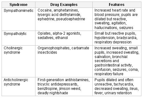
Knowledge of the principal toxidromes can be of considerable help to the clinician in raising suspicions of specific drug intoxications. These include lysergic acid diethylamide (LSD), cocaine, amphetamines, opioids, sedatives, ethanol, organophosphates, carbamate insecticides, jimson weed, deadly nightshade, ephedrine, pseudoephedrine, alpha-2 agonists, sedatives, first-generation antihistamines, tricyclic antidepressants, and benztropine. [Figure caption and citation for the preceding image starts]: The principal toxidromes, a constellation of features peculiar to certain classes of drugsTable created by G. Bryan Young, MD; used with permission [Citation ends].
Disorders mimicking coma
Psychogenic unresponsiveness and locked-in states may both be confused with coma. More importantly, locked-in state may even be confused with brain death.[40]
Psychogenic unresponsiveness may manifest as pseudocoma or nonepileptic seizures (pseudoseizures/psychogenic seizures). Although pseudocoma is uncommon, nonepileptic (pseudo) seizures make up about one third of admissions to epilepsy units.[41] There is often a background of psychosocial problems and abuse, as well as a lack of response to antiepileptic medications. Onset of psychogenic unresponsiveness in childhood or age >60 years is uncommon, but can occur. Most patients are women.[1]
Locked-in state is uncommon. Patients with locked-in state have preserved consciousness but impaired motor output. This includes patients with basis pontis lesions, severe polyneuropathies (e.g., Guillain-Barre syndrome, acute inflammatory demyelinating polyneuropathy), pharmacologic neuromuscular paralysis, and central pontine myelinolysis. Basis pontis lesions might be caused by an occlusion of the basilar artery, hypertensive hemorrhage, or central pontine myelinolysis. Central pontine myelinolysis typically occurs in systemically ill inpatients with sudden electrolyte disturbance. Pharmacologic paralysis most often occurs in the ICU or postsurgical recovery room, as a result of slow clearance or metabolism of neuromuscular blocking agents.
Use of this content is subject to our disclaimer