A thorough history and comprehensive clinical examination are critical to a clinical diagnosis, which is then confirmed by radiographs.
History
A history of trauma is almost always present. A fall on the outstretched hand (FOOSH) is often the cause and may occur from a simple slip or trip. This mechanism is more common in the older age groups.[14]Nellans KW, Kowalski E, Chung KC. The epidemiology of distal radius fractures. Hand Clin. 2012 May;28(2):113-25.
https://www.ncbi.nlm.nih.gov/pmc/articles/PMC3345129
http://www.ncbi.nlm.nih.gov/pubmed/22554654?tool=bestpractice.com
In such patients the possibility of osteoporosis as a predisposing factor for fractures should be considered. In the younger patient, there is often a history of a fall on the outstretched hand during a sporting activity, or a history of a vehicular trauma.[11]Court-Brown CM, Caesar B. Epidemiology of adult fractures: a review. Injury. 2006 Aug;37(8):691-7.
http://www.ncbi.nlm.nih.gov/pubmed/16814787?tool=bestpractice.com
[14]Nellans KW, Kowalski E, Chung KC. The epidemiology of distal radius fractures. Hand Clin. 2012 May;28(2):113-25.
https://www.ncbi.nlm.nih.gov/pmc/articles/PMC3345129
http://www.ncbi.nlm.nih.gov/pubmed/22554654?tool=bestpractice.com
Often the details of the exact cause/mechanism can be limited.
Physical examination
Nondisplaced fractures
Nondisplaced fractures usually present with localized swelling but no deformity. Tenderness over the distal radius is the hallmark.
Displaced fractures
Displaced fractures often present with the classic "dinner fork" deformity with dorsal angulation at the fracture site (resulting from dorsal displacement from falling on a pronated hand). The wrist is tender and range of motion is limited by pain.
The carpus, especially the scaphoid, should be carefully palpated in the anatomic snuff-box distal to the distal radius. Tenderness in this area is suggestive of a scaphoid fracture, either as an isolated injury or associated with concomitant fractures of the distal radius.[2]Rutgers M, Mudgal CS, Shin R. Combined fractures of the distal radius and scaphoid. J Hand Surg Eur Vol. 2008 Aug;33(4):478-83.
http://www.ncbi.nlm.nih.gov/pubmed/18687836?tool=bestpractice.com
Open fractures
Fractures of the distal radius can sometimes be associated with open wounds. Irrespective of age, an open wound usually suggests a high-energy injury.[6]Mudgal CS, Psenica J, Jupiter JB. Radiocarpal fracture-dislocation. J Hand Surg Br. 1999 Feb;24(1):92-8.
http://www.ncbi.nlm.nih.gov/pubmed/10190615?tool=bestpractice.com
In these fractures patients may experience numbness affecting the radial three digits, suggesting acute median nerve compression (carpal tunnel syndrome).[6]Mudgal CS, Psenica J, Jupiter JB. Radiocarpal fracture-dislocation. J Hand Surg Br. 1999 Feb;24(1):92-8.
http://www.ncbi.nlm.nih.gov/pubmed/10190615?tool=bestpractice.com
Numbness of the ulnar two digits, suggesting ulnar nerve compression, is less common. Signs of a vascular injury include lack of a palpable pulse, continued blood loss, and an expanding hematoma.[29]National Institute for Health and Care Excellence. Fractures (complex): assessment and management. Nov 2022 [internet publication].
https://www.nice.org.uk/guidance/ng37
The key clinical findings of compartment syndrome are pain out of proportion to the associated injury and pain on passive movement of the muscles of the involved compartments.[30]British Orthopaedic Association. BOAST - diagnosis and management of compartment syndrome of the limbs. Jul 2014 [internet publication].
https://www.boa.ac.uk/resources/boast-10-pdf.html
Pulses are normally present in compartment syndrome. Absent pulses are usually due to systemic hypotension, arterial occlusion, or vascular injury.[30]British Orthopaedic Association. BOAST - diagnosis and management of compartment syndrome of the limbs. Jul 2014 [internet publication].
https://www.boa.ac.uk/resources/boast-10-pdf.html
See Compartment syndrome of extremities.
Imaging
X-ray
An initial radiographic examination is required to make a conclusive diagnosis of a distal radius fracture.[31]Torabi M, Lenchik L, Beaman FD, et al; Expert Panel on Musculoskeletal Imaging. ACR appropriateness criteria: acute hand and wrist trauma. J Am Coll Radiol. 2019 May;16(5s):S7-17.
https://www.jacr.org/article/S1546-1440(19)30180-2/fulltext
http://www.ncbi.nlm.nih.gov/pubmed/31054760?tool=bestpractice.com
Typically posteroanterior, lateral, and carpal views should be obtained.[31]Torabi M, Lenchik L, Beaman FD, et al; Expert Panel on Musculoskeletal Imaging. ACR appropriateness criteria: acute hand and wrist trauma. J Am Coll Radiol. 2019 May;16(5s):S7-17.
https://www.jacr.org/article/S1546-1440(19)30180-2/fulltext
http://www.ncbi.nlm.nih.gov/pubmed/31054760?tool=bestpractice.com
Fractures may be minimal cracks, extra-articular fractures, or intra-articular fractures. Radiographs also suggest the degree of osteopenia, and may offer some information about the degree of articular involvement or comminution.
Although uncommon, combined injuries of the distal radius and scaphoid are seen. Scaphoid fractures can be missed and radiographs must be carefully examined for these fractures.[2]Rutgers M, Mudgal CS, Shin R. Combined fractures of the distal radius and scaphoid. J Hand Surg Eur Vol. 2008 Aug;33(4):478-83.
http://www.ncbi.nlm.nih.gov/pubmed/18687836?tool=bestpractice.com
In patients where there is pain or tenderness in the anatomic snuff-box a clinical diagnosis of a scaphoid fracture should be considered.
For fractures of the distal radius, radiographic features that suggest the need for surgical treatment include:[13]Jupiter JB. Complex articular fractures of the distal radius: classification and management. J Am Acad Orthop Surg. 1997 May;5(3):119-29.
http://www.ncbi.nlm.nih.gov/pubmed/10797214?tool=bestpractice.com
[32]Jupiter JB, Fernandez DL, Whipple TL, et al. Intra-articular fractures of the distal radius: contemporary perspectives. Instr Course Lect. 1998;47:191-202.
http://www.ncbi.nlm.nih.gov/pubmed/9571418?tool=bestpractice.com
[33]Jupiter JB, Ring D, Weitzel PP. Surgical treatment of redisplaced fractures of the distal radius in patients older than 60 years. J Hand Surg Am. 2002 Jul;27(4):714-23.
http://www.ncbi.nlm.nih.gov/pubmed/12132101?tool=bestpractice.com
[34]Johnson N, Leighton P, Distal Radius Fracture Delphi Study Group, et al. Defining displacement thresholds for surgical intervention for distal radius fractures - a Delphi study. PLoS One. 2019 Jan 8;14(1):e0210462.
https://journals.plos.org/plosone/article?id=10.1371/journal.pone.0210462
http://www.ncbi.nlm.nih.gov/pubmed/30620763?tool=bestpractice.com
[35]American Academy of Orthopaedic Surgeons. Management of distal radius fractures: evidence-based clinical practice guideline. Dec 2020 [internet publication].
https://www.aaos.org/globalassets/quality-and-practice-resources/distal-radius/drfcpg.pdf
Initial loss of radial length in excess of 15 mm
Angulation apex-volar of >20°
Postreduction radial shortening of >3 mm
Dorsal tilt of >10°
Intra-articular step-off of >2 mm.
Computed tomography (CT) scan
Do not order CT scans for children until physical exam has been completed and plain radiographs have been obtained. CT scans deliver a significant dose of radiation to a child, so their utility needs to be confirmed prior to ordering.[36]American Academy of Pediatrics - Section on Orthopaedics and the Pediatric Orthopaedic Society of North America. Five things physicians and patients should question. Choosing Wisely, an initiative of the ABIM Foundation. 2022 [internet publication].
https://web.archive.org/web/20230209025014/https://www.choosingwisely.org/societies/american-academy-of-pediatrics-section-on-orthopaedics-and-the-pediatric-orthopaedic-society-of-north-america
However, in patients where surgical fixation is considered, a CT scan of the distal radius is beneficial for the analysis of extra- and intra-articular fracture geometry and preoperative planning.[31]Torabi M, Lenchik L, Beaman FD, et al; Expert Panel on Musculoskeletal Imaging. ACR appropriateness criteria: acute hand and wrist trauma. J Am Coll Radiol. 2019 May;16(5s):S7-17.
https://www.jacr.org/article/S1546-1440(19)30180-2/fulltext
http://www.ncbi.nlm.nih.gov/pubmed/31054760?tool=bestpractice.com
A CT scan may also reveal any occult fractures around the carpus.
Magnetic resonance imaging (MRI)
Do not order MRI scans for children until physical exam has been completed and plain radiographs have been obtained. MRI scans may not be correctly interpreted without all of the necessary clinical information and frequently require sedation in a young child.[36]American Academy of Pediatrics - Section on Orthopaedics and the Pediatric Orthopaedic Society of North America. Five things physicians and patients should question. Choosing Wisely, an initiative of the ABIM Foundation. 2022 [internet publication].
https://web.archive.org/web/20230209025014/https://www.choosingwisely.org/societies/american-academy-of-pediatrics-section-on-orthopaedics-and-the-pediatric-orthopaedic-society-of-north-america
However, scaphoid fractures can be missed on x-ray; consider an MRI (without contrast) as first-line imaging if an occult fracture of the scaphoid is suspected.[37]National Institute for Health and Care Excellence. Fractures (non-complex): assessment and management. Feb 2016 [internet publication].
https://www.nice.org.uk/guidance/ng38
It may also be used to define associated ligamentous injuries.[38]Yin ZG, Zhang JB, Kan SL, et al. Diagnosing suspected scaphoid fractures: a systematic review and meta-analysis. Clin Orthop Relat Res. 2010 Mar;468(3):723-34.
https://journals.lww.com/clinorthop/Fulltext/2010/03000/Diagnosing_Suspected_Scaphoid_Fractures__A.12.aspx
http://www.ncbi.nlm.nih.gov/pubmed/19756904?tool=bestpractice.com
[39]American College of Radiology. ACR appropriateness criteria: chronic hand and wrist pain. 2023 [internet publication].
https://acsearch.acr.org/docs/69427/Narrative
Bone mineral density (BMD) / Dual-energy x-ray absorptiometry (DXA)
Assessment of bone mineral density should be performed as part of the follow-up in all patients over the age of 50 years.[16]British Orthopaedic Association and The British Society for Surgery of the Hand. Best practice for management of distal radius fracture. 2018 [internet publication].
https://www.bssh.ac.uk/professionals/management_of_distal_radial_fractures.aspx
DXA is considered the standard for measurement of bone density; the World Health Organization considers BMD measurements diagnostic for osteoporosis.[40]WHO Scientific Group on the Prevention and Management of Osteoporosis. Prevention and management of osteoporosis: report of a WHO scientific group. (WHO technical report series; 921.) Geneva, Switzerland; 2003.
https://apps.who.int/iris/handle/10665/42841
[Figure caption and citation for the preceding image starts]: Type A extra-articular fracture of the distal radius: lateral viewFrom the collection of Dr Chaitanya S. Mudgal [Citation ends]. [Figure caption and citation for the preceding image starts]: Type B (simple) intra-articular fracture of the distal radius: lateral viewFrom the collection of Dr Chaitanya S. Mudgal [Citation ends].
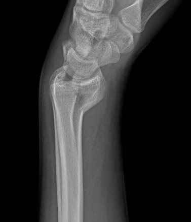 [Figure caption and citation for the preceding image starts]: Type C (complex) intra-articular fracture of the distal radius: lateral viewFrom the collection of Dr Chaitanya S. Mudgal [Citation ends].
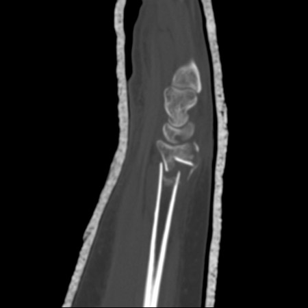
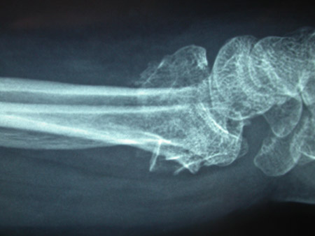 [Figure caption and citation for the preceding image starts]: CT scans of the wrist provide excellent detail to assess fracture geometry and articular involvement, as well as degree of comminutionFrom the collection of Dr Chaitanya S. Mudgal [Citation ends].