Images and videos
Images


Wrist fractures
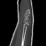
CT scans of the wrist provide excellent detail to assess fracture geometry and articular involvement, as well as degree of comminution
From the collection of Dr Chaitanya S. Mudgal
See this image in context in the following section/s:
Wrist fractures
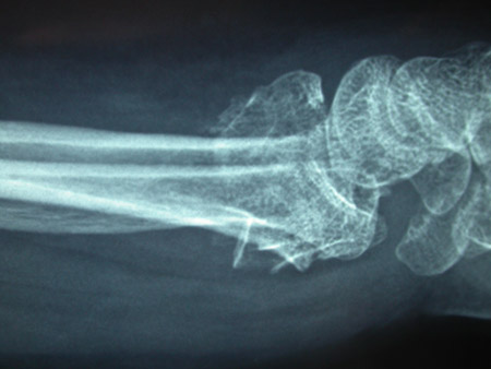
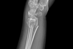
Type C (complex) intra-articular fracture of the distal radius: lateral view
From the collection of Dr Chaitanya S. Mudgal
See this image in context in the following section/s:
Wrist fractures
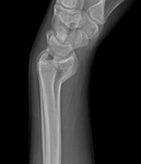
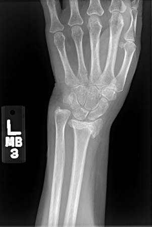
Posteroanterior radiograph showing malunion of the distal radius with significant shortening of the radius and relative lengthening of the ulna
From the collection of Dr Chaitanya S. Mudgal
See this image in context in the following section/s:

Wrist fractures
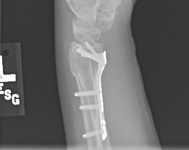
Plate fixation after open reduction with a volarly placed plate and screws
From the collection of Dr Chaitanya S. Mudgal
See this image in context in the following section/s:
Wrist fractures
Type A extra-articular fracture of the distal radius: lateral view
From the collection of Dr Chaitanya S. Mudgal
See this image in context in the following section/s:

Wrist fractures
Cast treatment of a distal radius fracture
From the collection of Dr Chaitanya S. Mudgal
See this image in context in the following section/s:

Wrist fractures
Type B (simple) intra-articular fracture of the distal radius: lateral view
From the collection of Dr Chaitanya S. Mudgal
See this image in context in the following section/s:
Use of this content is subject to our disclaimer