The goal of treatment of distal radius fractures is restoration of anatomy and recovery of hand, wrist, and forearm function.
Whether the patient is being treated in a cast or a splint or is waiting for a surgical fixation, it is important that they start rehabilitation of the hand at the earliest opportunity; patient education, reassurance, and pain control are essential during the initial visit.
The affected limb needs to be elevated. Active range of motion of the fingers and shoulder should begin during the first few days after injury. This is to help control edema in the hand, and to prevent stiffness in the metacarpophalangeal (MCP) and proximal interphalangeal joints, and frozen shoulder.[46]Miller LK, Jerosch-Herold C, Shepstone L. Effectiveness of edema management techniques for subacute hand edema: a systematic review. J Hand Ther. 2017 Oct-Dec;30(4):432-46.
https://www.jhandtherapy.org/article/S0894-1130(16)30228-9/fulltext
http://www.ncbi.nlm.nih.gov/pubmed/28807598?tool=bestpractice.com
[47]Itoi E, Arce G, Bain GI, et al. Shoulder stiffness: current concepts and concerns. Arthroscopy. 2016 Jul;32(7):1402-14.
http://www.ncbi.nlm.nih.gov/pubmed/27180923?tool=bestpractice.com
In all patients, following manual or surgical fracture reduction, if median nerve dysfunction is found to worsen or persists, a carpal tunnel release procedure should be performed urgently by an orthopedic surgeon.[48]Niver GE, Ilyas AM. Carpal tunnel syndrome after distal radius fracture. Orthop Clin North Am. 2012 Oct;43(4):521-7.
http://www.ncbi.nlm.nih.gov/pubmed/23026468?tool=bestpractice.com
[49]Brüske J, Niedźwiedź Z, Bednarski M, et al. Acute carpal tunnel syndrome after distal radius fractures - long term results of surgical treatment with decompression and external fixator application [in Polish]. Chir Narzadow Ruchu Ortop Pol. 2002;67(1):47-53.
http://www.ncbi.nlm.nih.gov/pubmed/12087674?tool=bestpractice.com
[50]Mack GR, McPherson SA, Lutz RB. Acute median neuropathy after wrist trauma. The role of emergent carpal tunnel release. Clin Orthop Relat Res. 1994 Mar;(300):141-6.
http://www.ncbi.nlm.nih.gov/pubmed/8131326?tool=bestpractice.com
Initiating a bone mineral density workup in the orthopedic clinic can improve osteoporosis evaluation and treatment rates following fragility fractures of the distal part of the radius.[16]British Orthopaedic Association and The British Society for Surgery of the Hand. Best practice for management of distal radius fracture. 2018 [internet publication].
https://www.bssh.ac.uk/professionals/management_of_distal_radial_fractures.aspx
[51]Rozental TD, Makhni EC, Day CS, et al. Improving evaluation and treatment for osteoporosis following distal radial fractures: a prospective randomized intervention. J Bone Joint Surg Am. 2008 May;90(5):953-61.
http://www.ncbi.nlm.nih.gov/pubmed/18451385?tool=bestpractice.com
Appropriate pain management is important, especially during rehabilitation; however, specific treatment varies widely depending on the patient, clinical presentation, method of treatment, and local treatment protocols. Opioid alternatives, both pharmacologic (e.g., local anesthetics, nonsteroidal anti-inflammatory drugs, acetaminophen) and nonpharmacologic (e.g., ice, elevation, compression, cognitive therapies) should be considered alongside opioid-sparing protocols when possible given the risks of opioid analgesics (adverse events, misuse, opioid use disorder, and diversion for nonmedical use).[35]American Academy of Orthopaedic Surgeons. Management of distal radius fractures: evidence-based clinical practice guideline. Dec 2020 [internet publication].
https://www.aaos.org/globalassets/quality-and-practice-resources/distal-radius/drfcpg.pdf
Ulnar styloid fractures often occur concurrently with distal radius fractures. They can usually be managed nonoperatively.[52]van Rossenberg LX, Beeres FJP, van Heijl M, et al. Operative versus non-operative treatment of ulnar styloid process base fractures: a systematic review and meta-analysis. Eur J Trauma Emerg Surg. 2024 Sep 13 [Epub ahead of print].
https://www.doi.org/10.1007/s00068-024-02660-2
http://www.ncbi.nlm.nih.gov/pubmed/39269646?tool=bestpractice.com
Initial treatment prior to definitive care
Closed fractures
For closed fractures, initial treatment is immobilization.
Typically, a short arm cast may be used with free MCP joints and not extending beyond the distal palmar flexion crease. Noncircumferential splints are often used acutely in the emergency department due to the risk of swelling.
If the fracture is displaced, the patient will require reduction or referral to a center where this can be performed. Postreduction radiographs should be obtained in the splint. If reduction is inadequate or unstable, an open reduction and fixation is likely to be necessary.
All fractures should have adequate follow-up care to ensure timely care. For unstable fractures and those requiring fixation, immediate consultation with an orthopedic surgeon should be obtained (or with a specialist hand surgeon if available).
Open fractures
Open fractures require urgent treatment with saline irrigation and debridement of the fracture and open wound, and removal of all devitalized tissue as well as foreign debris, prior to fixation.[53]Rupp M, Popp D, Alt V. Prevention of infection in open fractures: where are the pendulums now? Injury. 2020 May;51 Suppl 2:S57-63.
http://www.ncbi.nlm.nih.gov/pubmed/31679836?tool=bestpractice.com
Subsequently, if any delay in definitive treatment is anticipated, the fracture may be provisionally stabilized via a splint or an external fixator.[16]British Orthopaedic Association and The British Society for Surgery of the Hand. Best practice for management of distal radius fracture. 2018 [internet publication].
https://www.bssh.ac.uk/professionals/management_of_distal_radial_fractures.aspx
The American Academy of Orthopaedic Surgeons (AAOS) recommends that patients with open fractures are brought to the operating room for debridement and irrigation as soon as reasonable, and ideally within 24 hours post injury.[44]American Academy of Orthopaedic Surgeons. Prevention of surgical site infections after major extremity trauma: appropriate use criteria. Mar 2022 [internet publication].
https://www.aaos.org/globalassets/quality-and-practice-resources/dod/ssitrauma/ssitraumaauc.pdf
[54]American Academy of Orthopaedic Surgeons. Prevention of surgical site infections after major extremity trauma: evidence-based clinical practice guideline. Mar 2022 [internet publication].
https://www.aaos.org/globalassets/quality-and-practice-resources/dod/ssitrauma/ssitraumacpg.pdf
Preoperative antibiotics are recommended to prevent surgical site infections in operative treatment of open fractures.[44]American Academy of Orthopaedic Surgeons. Prevention of surgical site infections after major extremity trauma: appropriate use criteria. Mar 2022 [internet publication].
https://www.aaos.org/globalassets/quality-and-practice-resources/dod/ssitrauma/ssitraumaauc.pdf
[54]American Academy of Orthopaedic Surgeons. Prevention of surgical site infections after major extremity trauma: evidence-based clinical practice guideline. Mar 2022 [internet publication].
https://www.aaos.org/globalassets/quality-and-practice-resources/dod/ssitrauma/ssitraumacpg.pdf
In patients with major extremity trauma undergoing surgery, the AAOS strongly recommends antibiotic prophylaxis with systemic cefazolin or clindamycin, except for open fractures that are Type III (an open segmental fracture, or an open fracture with extensive soft tissue damage, or a traumatic amputation) and possibly Type II (laceration greater than 1 cm long without extensive soft tissue damage, flaps, or avulsions), for which additional gram-negative coverage (e.g., piperacillin/tazobactam) is preferred.[44]American Academy of Orthopaedic Surgeons. Prevention of surgical site infections after major extremity trauma: appropriate use criteria. Mar 2022 [internet publication].
https://www.aaos.org/globalassets/quality-and-practice-resources/dod/ssitrauma/ssitraumaauc.pdf
[54]American Academy of Orthopaedic Surgeons. Prevention of surgical site infections after major extremity trauma: evidence-based clinical practice guideline. Mar 2022 [internet publication].
https://www.aaos.org/globalassets/quality-and-practice-resources/dod/ssitrauma/ssitraumacpg.pdf
[55]Gustilo RB, Anderson JT. Prevention of infection in the treatment of one thousand and twenty-five open fractures of long bones: retrospective and prospective analyses. J Bone Joint Surg Am. 1976 Jun;58(4):453-8.
http://www.ncbi.nlm.nih.gov/pubmed/773941?tool=bestpractice.com
However, local sensitivities and protocols should be followed for antibiotic selection. In patients with major extremity trauma undergoing surgery, local antibiotic prophylactic strategies, such as vancomycin powder, tobramycin-impregnated beads, or gentamicin-covered nails, may be beneficial, when available.[44]American Academy of Orthopaedic Surgeons. Prevention of surgical site infections after major extremity trauma: appropriate use criteria. Mar 2022 [internet publication].
https://www.aaos.org/globalassets/quality-and-practice-resources/dod/ssitrauma/ssitraumaauc.pdf
[54]American Academy of Orthopaedic Surgeons. Prevention of surgical site infections after major extremity trauma: evidence-based clinical practice guideline. Mar 2022 [internet publication].
https://www.aaos.org/globalassets/quality-and-practice-resources/dod/ssitrauma/ssitraumacpg.pdf
One study suggests that open fractures should be debrided within 6 hours of injury.[56]Prodromidis AD, Charalambous CP. The 6-hour rule for surgical debridement of open tibial fractures: a systematic review and meta-analysis of infection and nonunion Rates. J Orthop Trauma. 2016 Jul;30(7):397-402.
http://www.ncbi.nlm.nih.gov/pubmed/26978135?tool=bestpractice.com
Another prospective study concludes that time to irrigation and debridement does not affect the development of local infections, provided it is performed within 24 hours of arrival to the emergency department.[57]Srour M, Inaba K, Okoye O, et al. Prospective evaluation of treatment of open fractures: effect of time to irrigation and debridement. JAMA Surg. 2015 Apr;150(4):332-6.
https://jamanetwork.com/journals/jamasurgery/fullarticle/2108746
http://www.ncbi.nlm.nih.gov/pubmed/25692391?tool=bestpractice.com
One systematic review reports that early debridement of open fractures by an experienced team within 24 hours is adequate.[53]Rupp M, Popp D, Alt V. Prevention of infection in open fractures: where are the pendulums now? Injury. 2020 May;51 Suppl 2:S57-63.
http://www.ncbi.nlm.nih.gov/pubmed/31679836?tool=bestpractice.com
Should contamination be a concern, it is prudent to perform irrigation and debridement, and to provisionally stabilize the fracture via an external fixator.[44]American Academy of Orthopaedic Surgeons. Prevention of surgical site infections after major extremity trauma: appropriate use criteria. Mar 2022 [internet publication].
https://www.aaos.org/globalassets/quality-and-practice-resources/dod/ssitrauma/ssitraumaauc.pdf
[54]American Academy of Orthopaedic Surgeons. Prevention of surgical site infections after major extremity trauma: evidence-based clinical practice guideline. Mar 2022 [internet publication].
https://www.aaos.org/globalassets/quality-and-practice-resources/dod/ssitrauma/ssitraumacpg.pdf
For treatment of infections, antibiotics should be administered according to the type of open injury and severity of contamination at the time of diagnosis.[44]American Academy of Orthopaedic Surgeons. Prevention of surgical site infections after major extremity trauma: appropriate use criteria. Mar 2022 [internet publication].
https://www.aaos.org/globalassets/quality-and-practice-resources/dod/ssitrauma/ssitraumaauc.pdf
[53]Rupp M, Popp D, Alt V. Prevention of infection in open fractures: where are the pendulums now? Injury. 2020 May;51 Suppl 2:S57-63.
http://www.ncbi.nlm.nih.gov/pubmed/31679836?tool=bestpractice.com
However, local sensitivities and protocols should be followed for antibiotic selection.
After the infection is controlled, a formal open reduction and internal fixation may be performed at a later stage.[54]American Academy of Orthopaedic Surgeons. Prevention of surgical site infections after major extremity trauma: evidence-based clinical practice guideline. Mar 2022 [internet publication].
https://www.aaos.org/globalassets/quality-and-practice-resources/dod/ssitrauma/ssitraumacpg.pdf
Monitoring for median nerve function should be maintained throughout the postoperative period.
Nondisplaced fractures of distal radius
Patients with nondisplaced fractures of the distal radius usually have sustained low-energy injuries and are largely comfortable once the wrist is immobilized.
The choice of immobilization may vary from a cast applied by the surgeon or a cast technician, to a custom-made splint from an occupational therapist.
Short arm casts made of plaster of Paris or fiberglass are applied distal to the elbow and in nondisplaced fractures maintain the position of the wrist at neutral.[16]British Orthopaedic Association and The British Society for Surgery of the Hand. Best practice for management of distal radius fracture. 2018 [internet publication].
https://www.bssh.ac.uk/professionals/management_of_distal_radial_fractures.aspx
The thumb is free and the cast terminates at the level of the distal palmar flexion crease. This allows free motion of the MCP joints, thus maintaining digital mobility as the fracture heals, and minimizes post-traumatic stiffness.
Casts should be well fitting, and well padded to avoid any pressure effects, and the patient must be alerted to the possibility of needing a cast change. Cast changes may be necessary if the cast gets loose as the initial post-traumatic swelling reduces. Cast immobilization for a period of 3-4 weeks is safe.[16]British Orthopaedic Association and The British Society for Surgery of the Hand. Best practice for management of distal radius fracture. 2018 [internet publication].
https://www.bssh.ac.uk/professionals/management_of_distal_radial_fractures.aspx
[58]Bentohami A, van Delft EAK, Vermeulen J, et al. Non- or minimally displaced distal radial fractures in adult patients: three weeks versus five weeks of cast immobilization - a randomized controlled trial. J Wrist Surg. 2019 Feb;8(1):43-8.
https://www.ncbi.nlm.nih.gov/pmc/articles/PMC6358449
http://www.ncbi.nlm.nih.gov/pubmed/30723601?tool=bestpractice.com
[59]Delft EAKV, Gelder TGV, Vries R, et al. Duration of cast immobilization in distal radial fractures: a systematic review. J Wrist Surg. 2019 Oct;8(5):430-8.
http://www.ncbi.nlm.nih.gov/pubmed/31579555?tool=bestpractice.com
Alternatively, in patients unable to tolerate casts or unwilling to wear a cast, or in patients who have an incomplete fracture of the distal radius, a forearm-based splint holding the wrist at neutral may be used.[60]Roth KM, Blazar PE, Earp BE, et al. Incidence of displacement after nondisplaced distal radial fractures in adults. J Bone Joint Surg Am. 2013 Aug 7;95(15):1398-402.
http://www.ncbi.nlm.nih.gov/pubmed/23925744?tool=bestpractice.com
Splints are custom-made by occupational therapists, and can be custom-molded to the patient's anatomy. As swelling reduces, modification to fit the changing dimensions of the patient's limb may be necessary.
Patients with nondisplaced fractures or incomplete crack fractures of the radius must be cautioned about the possibility of spontaneous rupture of the extensor pollicis longus (EPL) tendon. This is a rare complication with an incidence of 5% or less, and tends to occur within the first 12 weeks after injury and is usually preceded by increasing pain over the dorsal aspect of the distal radius.[61]Roth KM, Blazar PE, Earp BE, et al. Incidence of extensor pollicis longus tendon rupture after nondisplaced distal radius fractures. J Hand Surg Am. 2012 May;37(5):942-7.
http://www.ncbi.nlm.nih.gov/pubmed/22463927?tool=bestpractice.com
Not all EPL ruptures are symptomatic and not all necessarily need to be treated. [Figure caption and citation for the preceding image starts]: Cast treatment of a distal radius fractureFrom the collection of Dr Chaitanya S. Mudgal [Citation ends].
Closed displaced fracture of distal radius
Restoration of anatomy is essential in an effort to maximize functional outcome. In the initial stages this may be achieved by a manipulative reduction, and thereafter formal treatment is planned.
Analysis of fracture geometry is aided by post-reduction radiographs, and if necessary a computed tomography scan may be obtained. These radiographic investigations offer clues to the stability or instability of the fracture and help guide its definitive treatment.
Manipulative reduction in the acute setting is most often performed using a hematoma block (instillation of a local anesthetic within the fracture hematoma), and distraction of the fracture site aided by finger traps.[62]Myderrizi N, Mema B. The hematoma block an effective alternative for fracture reduction in distal radius fractures. Med Arh. 2011;65(4):239-42.
http://www.ncbi.nlm.nih.gov/pubmed/21950232?tool=bestpractice.com
[63]Tseng PT, Leu TH, Chen YW, et al. Hematoma block or procedural sedation and analgesia, which is the most effective method of anesthesia in reduction of displaced distal radius fracture? J Orthop Surg Res. 2018 Mar 27;13(1):62.
https://josr-online.biomedcentral.com/articles/10.1186/s13018-018-0772-7
http://www.ncbi.nlm.nih.gov/pubmed/29580286?tool=bestpractice.com
[64]Søsborg-Würtz H, Corap Gellert S, Ladeby Erichsen J, et al. Closed reduction of distal radius fractures: a systematic review and meta-analysis. EFORT Open Rev. 2018 Apr;3(4):114-20.
https://eor.bioscientifica.com/view/journals/eor/3/4/2058-5241.3.170063.xml
http://www.ncbi.nlm.nih.gov/pubmed/29780618?tool=bestpractice.com
Diffusion of the anesthetic volarly around the median and ulnar nerves may occur, and patients should be reassured that digital numbness is to be expected. Conscious sedation may be used for reductions in the emergency department. Different fracture geometries require different reduction techniques; for example, a dorsally angulated fracture can be reduced by applying dorsal pressure to the distal fragment to "milk" it back into position.
Adequate reduction is verified by palpation for step-offs along the dorsal and radial surfaces. The fracture is then held in its reduced position in a well-molded splint. Postreduction radiographs should be obtained with the splint in place.
If the patient is an unsuitable candidate for prolonged casting or inadequate reduction is observed on imaging following manual reduction, surgical reduction and fixation should be considered.
The decision about whether a surgical intervention is warranted should be discussed between the patient and surgeon. Strong evidence suggests no significant difference in radiographic or patient-reported outcomes between fixation techniques for complete articular or unstable distal radius fractures, although volar locking plates lead to earlier recovery of function in the short term (3-6 months), and outcomes equalize within 1 year of injury.[35]American Academy of Orthopaedic Surgeons. Management of distal radius fractures: evidence-based clinical practice guideline. Dec 2020 [internet publication].
https://www.aaos.org/globalassets/quality-and-practice-resources/distal-radius/drfcpg.pdf
[65]Selles CA, Mulders MAM, Winkelhagen J, et al; VIPAR Collaborators. Volar plate fixation versus cast immobilization in acceptably reduced intra-articular distal radial fractures: a randomized controlled trial. J Bone Joint Surg Am. 2021 Nov 3;103(21):1963-9.
http://www.ncbi.nlm.nih.gov/pubmed/34314402?tool=bestpractice.com
One randomized controlled trial involving 90 patients (46 in the nonoperative group and 44 in the operative group) found that 28% of nonoperatively managed patients had a subsequent surgical procedure.[65]Selles CA, Mulders MAM, Winkelhagen J, et al; VIPAR Collaborators. Volar plate fixation versus cast immobilization in acceptably reduced intra-articular distal radial fractures: a randomized controlled trial. J Bone Joint Surg Am. 2021 Nov 3;103(21):1963-9.
http://www.ncbi.nlm.nih.gov/pubmed/34314402?tool=bestpractice.com
Another randomized controlled trial reported that patients with an acceptably reduced extra-articular distal radial fracture treated with open reduction and volar plate fixation have better functional outcomes after 12 months compared with nonoperatively managed patients. A total of 42% of nonoperatively managed patients had a subsequent surgical procedure.[66]Mulders MAM, Walenkamp MMJ, van Dieren S, et al. Volar plate fixation versus plaster immobilization in acceptably reduced extra-articular distal radial fractures: a multicenter randomized controlled trial. J Bone Joint Surg Am. 2019 May 1;101(9):787-96.
http://www.ncbi.nlm.nih.gov/pubmed/31045666?tool=bestpractice.com
Moderate evidence supports that for nongeriatric patients (most commonly defined in studies as under 65 years of age), operative treatment for fractures with post-reduction radial shortening >3 mm, dorsal tilt >10°, or intra-articular displacement or step off >2 mm leads to improved radiographic and patient-reported outcomes.[35]American Academy of Orthopaedic Surgeons. Management of distal radius fractures: evidence-based clinical practice guideline. Dec 2020 [internet publication].
https://www.aaos.org/globalassets/quality-and-practice-resources/distal-radius/drfcpg.pdf
Strong evidence suggests that operative treatment for geriatric patients (most commonly defined in studies as 65 years of age and older) does not lead to improved long-term patient-reported outcomes compared with nonoperative treatment.[35]American Academy of Orthopaedic Surgeons. Management of distal radius fractures: evidence-based clinical practice guideline. Dec 2020 [internet publication].
https://www.aaos.org/globalassets/quality-and-practice-resources/distal-radius/drfcpg.pdf
Following volar plate fixation, patients can be safely treated with a pressure bandage.[67]Poiset S, Abboudi J, Gallant G, et al. Splinting after distal radius fracture fixation: a prospective cohort analysis of postoperative plaster splint versus soft dressing. J Wrist Surg. 2019 Dec;8(6):452-5.
http://www.ncbi.nlm.nih.gov/pubmed/31815058?tool=bestpractice.com
Some surgeons prefer a cast for pain management. However, this period should not be longer than 3 weeks.[68]Watson N, Haines T, Tran P, et al. A comparison of the effect of one, three, or six weeks of immobilization on function and pain after open reduction and internal fixation of distal radial fractures in adults: a randomized controlled trial. J Bone Joint Surg Am. 2018 Jul 5;100(13):1118-25.
http://www.ncbi.nlm.nih.gov/pubmed/29975268?tool=bestpractice.com
In older patients with low functional demands, the presence of deformity does not preclude good functional outcomes.[69]Gehrmann SV, Windolf J, Kaufmann RA, et al. Distal radius fracture management in elderly patients: a literature review. J Hand Surg Am. 2008 Mar;33(3):421-9.
http://www.ncbi.nlm.nih.gov/pubmed/18343302?tool=bestpractice.com
Monitoring for median nerve function should be maintained throughout the postoperative period. [Figure caption and citation for the preceding image starts]: Plate fixation after open reduction with a volarly placed plate and screwsFrom the collection of Dr Chaitanya S. Mudgal [Citation ends].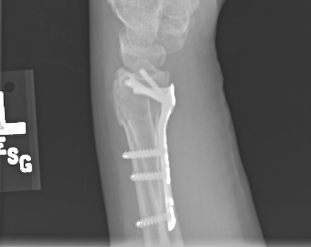 There is no apparent treatment benefit for the use of wrist arthroscopy at the time of distal radius fracture fixation.[35]American Academy of Orthopaedic Surgeons. Management of distal radius fractures: evidence-based clinical practice guideline. Dec 2020 [internet publication].
https://www.aaos.org/globalassets/quality-and-practice-resources/distal-radius/drfcpg.pdf
[70]Yamazaki H, Uchiyama S, Komatsu M, et al. Arthroscopic assistance does not improve the functional or radiographic outcome of unstable intra-articular distal radial fractures treated with a volar locking plate: a randomised controlled trial. Bone Joint J. 2015 Jul;97-B(7):957-62.
https://online.boneandjoint.org.uk/doi/full/10.1302/0301-620X.97B7.35354
http://www.ncbi.nlm.nih.gov/pubmed/26130352?tool=bestpractice.com
[71]Selles CA, Reerds STH, Roukema G, et al. Relationship between plate removal and Soong grading following surgery for fractured distal radius. J Hand Surg Eur Vol. 2018 Feb;43(2):137-41.
https://journals.sagepub.com/doi/10.1177/1753193417726636
http://www.ncbi.nlm.nih.gov/pubmed/28825371?tool=bestpractice.com
There is no apparent treatment benefit for the use of wrist arthroscopy at the time of distal radius fracture fixation.[35]American Academy of Orthopaedic Surgeons. Management of distal radius fractures: evidence-based clinical practice guideline. Dec 2020 [internet publication].
https://www.aaos.org/globalassets/quality-and-practice-resources/distal-radius/drfcpg.pdf
[70]Yamazaki H, Uchiyama S, Komatsu M, et al. Arthroscopic assistance does not improve the functional or radiographic outcome of unstable intra-articular distal radial fractures treated with a volar locking plate: a randomised controlled trial. Bone Joint J. 2015 Jul;97-B(7):957-62.
https://online.boneandjoint.org.uk/doi/full/10.1302/0301-620X.97B7.35354
http://www.ncbi.nlm.nih.gov/pubmed/26130352?tool=bestpractice.com
[71]Selles CA, Reerds STH, Roukema G, et al. Relationship between plate removal and Soong grading following surgery for fractured distal radius. J Hand Surg Eur Vol. 2018 Feb;43(2):137-41.
https://journals.sagepub.com/doi/10.1177/1753193417726636
http://www.ncbi.nlm.nih.gov/pubmed/28825371?tool=bestpractice.com
Open displaced fracture of distal radius
Open distal radius fractures are rare injuries.[72]Iorio ML, Harper CM, Rozental TD. Open distal radius fractures: timing and strategies for surgical management. Hand Clin. 2018 Feb;34(1):33-40.
http://www.ncbi.nlm.nih.gov/pubmed/29169595?tool=bestpractice.com
In most situations, open fractures constitute a surgical emergency and operative treatment at the earliest possible opportunity is the preferred method of management.
In patients who may have significant comorbidities or other injuries, a thorough evaluation of the risk of urgent operative intervention is critical, and is guided by a comprehensive discussion with other medical teams as well as the anesthesia team.
Fractures are often provisionally reduced in the emergency department in anticipation of delay in getting the patient into the operating room. This helps to reduce deformity and soft-tissue swelling, and may relieve any symptoms of nerve compression. Open reduction and internal fixation is performed in the operating room. Thorough saline irrigation and debridement of the fracture and the open wound is required prior to fixation.[44]American Academy of Orthopaedic Surgeons. Prevention of surgical site infections after major extremity trauma: appropriate use criteria. Mar 2022 [internet publication].
https://www.aaos.org/globalassets/quality-and-practice-resources/dod/ssitrauma/ssitraumaauc.pdf
[53]Rupp M, Popp D, Alt V. Prevention of infection in open fractures: where are the pendulums now? Injury. 2020 May;51 Suppl 2:S57-63.
http://www.ncbi.nlm.nih.gov/pubmed/31679836?tool=bestpractice.com
[54]American Academy of Orthopaedic Surgeons. Prevention of surgical site infections after major extremity trauma: evidence-based clinical practice guideline. Mar 2022 [internet publication].
https://www.aaos.org/globalassets/quality-and-practice-resources/dod/ssitrauma/ssitraumacpg.pdf
Should contamination be a concern and if there is a high concern for infection, internal fixation may be delayed or external fixation may be utilized as definitive treatment.[44]American Academy of Orthopaedic Surgeons. Prevention of surgical site infections after major extremity trauma: appropriate use criteria. Mar 2022 [internet publication].
https://www.aaos.org/globalassets/quality-and-practice-resources/dod/ssitrauma/ssitraumaauc.pdf
[54]American Academy of Orthopaedic Surgeons. Prevention of surgical site infections after major extremity trauma: evidence-based clinical practice guideline. Mar 2022 [internet publication].
https://www.aaos.org/globalassets/quality-and-practice-resources/dod/ssitrauma/ssitraumacpg.pdf
[72]Iorio ML, Harper CM, Rozental TD. Open distal radius fractures: timing and strategies for surgical management. Hand Clin. 2018 Feb;34(1):33-40.
http://www.ncbi.nlm.nih.gov/pubmed/29169595?tool=bestpractice.com
Ongoing antibiotic therapy should be administered according to the type of open injury and severity of contamination at the time of diagnosis.[44]American Academy of Orthopaedic Surgeons. Prevention of surgical site infections after major extremity trauma: appropriate use criteria. Mar 2022 [internet publication].
https://www.aaos.org/globalassets/quality-and-practice-resources/dod/ssitrauma/ssitraumaauc.pdf
In most centers, the specific regimen is best decided upon with infectious disease colleagues, taking local sensitivities into account.
Tetanus prophylaxis may need to be considered, depending on the patient's tetanus vaccination history.[73]Centers for Disease Control and Prevention. Clinical guidance for wound management to prevent tetanus. Aug 2024 [internet publication].
https://www.cdc.gov/tetanus/hcp/clinical-guidance
Most open fractures are high-energy injuries and there is a low threshold to perform concomitant carpal tunnel release. Such injuries may be accompanied by significant soft-tissue trauma. It is not uncommon for these patients to present with very swollen and tense forearms. In these patients it is mandatory to evaluate for forearm compartment syndrome. If there are obvious signs and symptoms of compartment syndrome, a clinical diagnosis is established and surgical fasciotomy is performed.[74]Duckworth AD, Mitchell SE, Molyneux SG, et al. Acute compartment syndrome of the forearm. J Bone Joint Surg Am. 2012 May 16;94(10):e63.
http://www.ncbi.nlm.nih.gov/pubmed/22617929?tool=bestpractice.com
This is performed in the emergency department, and a compartment pressure >30 mmHg is diagnostic of a compartment syndrome (if the patient is normotensive). This is a surgical emergency and urgent fasciotomy, combined with open reduction and fixation of the fracture as well as release of the carpal tunnel, is critical to minimize adverse long-term effects. See Compartment syndrome of extremities.
Nondisplaced fracture of the scaphoid
Isolated nondisplaced scaphoid fractures can be treated nonoperatively in most patients, with high union rates and good clinical outcomes.[75]Clementson M, Jørgsholm P, Besjakov J, et al. Conservative treatment versus arthroscopic-assisted screw fixation of scaphoid waist fractures - a randomized trial with minimum 4-year follow-up. J Hand Surg Am. 2015 Jul;40(7):1341-8.
http://www.ncbi.nlm.nih.gov/pubmed/25913660?tool=bestpractice.com
[76]Fowler JR, Hughes TB. Scaphoid fractures. Clin Sports Med. 2015 Jan;34(1):37-50.
http://www.ncbi.nlm.nih.gov/pubmed/25455395?tool=bestpractice.com
Patients are placed in a forearm-based cast without incorporating the thumb. There is no universal consensus on the duration of casting, but usually the cast is maintained for a total of 8-12 weeks or until the fracture is healed.[77]Buijze GA, Goslings JC, Rhemrev SJ, et al; CAST Trial Collaboration. Cast immobilization with and without immobilization of the thumb for nondisplaced and minimally displaced scaphoid waist fractures: a multicenter, randomized, controlled trial. J Hand Surg Am. 2014 Apr;39(4):621-7.
http://www.ncbi.nlm.nih.gov/pubmed/24582846?tool=bestpractice.com
Patients with proximal pole fractures, those with fracture displacement, or those who are unwilling to accept the protracted duration of casting are considered candidates for percutaneous screw fixation or for open reduction and internal fixation of the scaphoid.[78]Shen L, Tang J, Luo C, et al. Comparison of operative and non-operative treatment of acute undisplaced or minimally-displaced scaphoid fractures: a meta-analysis of randomized controlled trials. PLoS One. 2015 May 5;10(5):e0125247.
https://journals.plos.org/plosone/article?id=10.1371/journal.pone.0125247
http://www.ncbi.nlm.nih.gov/pubmed/25942316?tool=bestpractice.com
[79]Alnaeem H, Aldekhayel S, Kanevsky J, et al. A systematic review and meta-analysis examining the differences between nonsurgical management and percutaneous fixation of minimally and nondisplaced scaphoid fractures. J Hand Surg Am. 2016 Dec;41(12):1135-44;e1.
http://www.ncbi.nlm.nih.gov/pubmed/27707564?tool=bestpractice.com
Fractures of the distal radius with associated injury to carpus or carpal ligaments
Concomitant carpal bone, ligament injuries, or triangular fibrocartilage complex (TFCC) lesions may adversely impact outcome in patients with fractures of the distal radius.[2]Rutgers M, Mudgal CS, Shin R. Combined fractures of the distal radius and scaphoid. J Hand Surg Eur Vol. 2008 Aug;33(4):478-83.
http://www.ncbi.nlm.nih.gov/pubmed/18687836?tool=bestpractice.com
[80]Fernandez DL, Jupiter JB. Fractures of the distal radius. New York, NY: Springer-Verlag; 2002:41-7.[81]Young BT, Rayan GM. Outcome following nonoperative treatment of displaced distal radius fractures in low-demand patients older than 60 years. J Hand Surg Am. 2000 Jan;25(1):19-28.
http://www.ncbi.nlm.nih.gov/pubmed/10642469?tool=bestpractice.com
Fracture patterns in which the fracture line of the distal radius exits on the scapholunate crest of the articular surface are particularly prone to injuries of the scapholunate interosseous ligament.
Evidence suggests that distal radius fracture without concomitant ligament injury is rare. Arthroscopic studies report the incidence of concomitant scapholunate ligament (SL) and TFCC injury as 78% and 54% respectively in patients with distal radius fracture.[82]Mrkonjic A, Lindau T, Geijer M, et al. Arthroscopically diagnosed scapholunate ligament injuries associated with distal radial fractures: a 13- to 15-year follow-up. J Hand Surg Am. 2015 Jun;40(6):1077-82.
http://www.ncbi.nlm.nih.gov/pubmed/25936737?tool=bestpractice.com
[83]Mrkonjic A, Geijer M, Lindau T, et al. The natural course of traumatic triangular fibrocartilage complex tears in distal radial fractures: a 13-15 year follow-up of arthroscopically diagnosed but untreated injuries. J Hand Surg Am. 2012 Aug;37(8):1555-60.
http://www.ncbi.nlm.nih.gov/pubmed/22835585?tool=bestpractice.com
[84]Selles CA, Mulders MAM, Colaris JW, et al. Arthroscopic debridement does not enhance surgical treatment of intra-articular distal radius fractures: a randomized controlled trial. J Hand Surg Eur Vol. 2020 May;45(4):327-32.
http://www.ncbi.nlm.nih.gov/pubmed/31686586?tool=bestpractice.com
Two prospective 13- to 15-year follow-up studies of patients with untreated complete (grade 3) or partial (grade 1 or 2) SL tears and TFCC tears associated with displaced distal radius fracture found no major differences in the subjective, objective, or radiographic outcomes for patients with SL injuries. However, none of the patients had a grade 4 tear, and only one patient with a TFCC tear required an operation for painful instability since the fracture.[82]Mrkonjic A, Lindau T, Geijer M, et al. Arthroscopically diagnosed scapholunate ligament injuries associated with distal radial fractures: a 13- to 15-year follow-up. J Hand Surg Am. 2015 Jun;40(6):1077-82.
http://www.ncbi.nlm.nih.gov/pubmed/25936737?tool=bestpractice.com
[83]Mrkonjic A, Geijer M, Lindau T, et al. The natural course of traumatic triangular fibrocartilage complex tears in distal radial fractures: a 13-15 year follow-up of arthroscopically diagnosed but untreated injuries. J Hand Surg Am. 2012 Aug;37(8):1555-60.
http://www.ncbi.nlm.nih.gov/pubmed/22835585?tool=bestpractice.com
The TFCC tear study concluded that there was insufficient evidence that TFCC tear at the time of distal radius fracture would influence the subjective long-term outcome.[83]Mrkonjic A, Geijer M, Lindau T, et al. The natural course of traumatic triangular fibrocartilage complex tears in distal radial fractures: a 13-15 year follow-up of arthroscopically diagnosed but untreated injuries. J Hand Surg Am. 2012 Aug;37(8):1555-60.
http://www.ncbi.nlm.nih.gov/pubmed/22835585?tool=bestpractice.com