Complications
Fractures that are untreated, or those that are treated with nonoperative methods with a loss of position, develop a malunion. Close radiographic follow-up of a nonoperatively treated fracture is mandatory within the first 6 weeks to avoid a malunion. All malunions result in radiographic deformity. However, the cosmetic deformity may often be modest and functionally a malunion may not always require treatment. Patients with malunion and pain usually need treatment.[92][Figure caption and citation for the preceding image starts]: Posteroanterior radiograph showing malunion of the distal radius with significant shortening of the radius and relative lengthening of the ulnaFrom the collection of Dr Chaitanya S. Mudgal [Citation ends].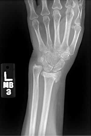
Stiffness of the joints in the hand is seen commonly after this fracture and occurs irrespective of method of treatment. It is essential to avoid certain features of treatment that may increase the possibility of this occurrence. Casts or splints that do not allow mobilization of the metacarpophalangeal and interphalangeal joints will lead to stiffness. An external fixator that is applied inappropriately may also have the same result.
Patient education in the importance of early digital motion, elevation, and edema control with compliance with occupational therapy will minimize this occurrence.[92]
CRPS is now also known as sympathetically mediated pain; other synonymous terms include: Sudeck atrophy osteodystrophy; causalgia; algodystrophy; and reflex sympathetic dystrophy.
CRPS is characterized by pain that may appear to be out of proportion to the injury and usually manifests within the first few weeks after the fracture. It is usually accompanied by stiffness and signs of sympathetic dysfunction including swelling, color changes, temperature changes, and changes in patterns of hair and nail growth. Patients with this condition are often best managed in conjunction with the pain management service. Aggressive physical therapy, tricyclic antidepressants, and stellate ganglion blocks are some options for treatment.[92]
With persisting osteoporosis, patients are at risk of further fragility fractures of the wrist, femoral neck, and vertebral bodies.
These are perhaps the most common long-term complication of distal radius fractures, irrespective of method of treatment. In most instances, ulnar-sided wrist pain not associated with a distal radius malunion will resolve over a period of 9-12 months and require little active intervention. Ulnar styloid nonunions are a radiographic diagnosis and cause few, if any, long-term symptoms.[94]
Most commonly this is seen after malunited intra-articular fractures of the distal radius, and may affect the radiocarpal joint or the distal radioulnar joint or both. Symptoms usually consist of pain during normal daily activity. Radiographic arthritis does not require treatment in the absence of symptoms. Treatment may include splinting, intra-articular corticosteroid injections, and surgical reconstruction.[92]
CTS can occur in the acute setting after displaced fractures or may be seen following malunion of distal radius fractures. One matched case control study reported that patients with open fracture, AO C-type distal radial fracture (DRFx), or with any intervening surgical procedure are at increased risk of developing acute CTS after DRFx post open reduction internal fixation.[91]
At the initial evaluation, a thorough neurologic examination must be performed prior to instillation of the hematoma block. In most instances, following an expeditious closed manipulation, median nerve symptoms resolve. If the symptoms do not resolve or become worse within 24-36 hours after manipulation, the patient will require surgical release of the carpal tunnel accompanied by fixation of the fracture. CTS associated with a malunited distal radius fracture usually requires surgical release of the carpal tunnel.[2][6][92]
This rare complication is seen most commonly in patients with completely nondisplaced fractures or crack fractures of the distal radius. It usually occurs within the first 12 weeks after the injury, and patients with such fractures should be cautioned regarding this rare but distinct possibility.[61]
The cause of the rupture is not clear and is largely believed to be due to attrition from alteration of blood supply to the tendon within its compartment over the distal radius. Patients who have increasing pain over the distal radius and with thumb motion, despite adequate immobilization, may be at risk for an EPL rupture. Not all patients with an EPL rupture need treatment. Loss of interphalangeal joint extension with a lag may cause functional difficulties, which may require tendon transfer to replace the ruptured EPL.[92]
This is a complication of treatment and seen after internal fixation of distal radius fractures with a plate placed volarly. Other tendons such as the flexor digitorum profundus of the index finger can also rupture. The cause of the rupture is thought to be attrition of tendons as they come in contact with a prominent plate.
Use of this content is subject to our disclaimer