Images and videos
Images


Aortic stenosis
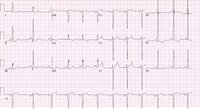
ECG showing changes associated with left ventricular hypertrophy
From the collection of Melanie Everitt MD, Heart Failure & Transplantation Program, Primary Children's Medical Center, Salt Lake City, UT; used with permission
See this image in context in the following section/s:

Aortic stenosis
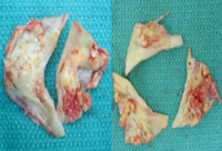
Bicuspid and trileaflet aortic valves with severe calcification following surgical excision
From the collection of David Liff, MD, Emory University Hospital; used with permission
See this image in context in the following section/s:
Aortic stenosis
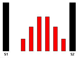
The systolic crescendo-decrescendo murmur of aortic stenosis
From the collection of David Liff, MD, Emory University Hospital; used with permission
See this image in context in the following section/s:
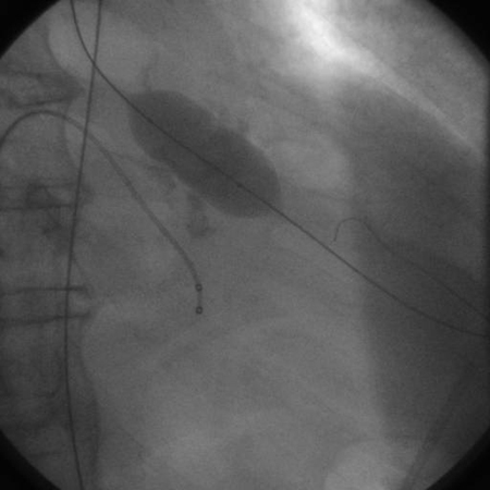
Aortic stenosis
Balloon valvuloplasty fluoroscopy film that demonstrates valvuloplasty balloon inflated across a calcified aortic valve
From the collection of David Liff, MD, Emory University Hospital; used with permission
See this image in context in the following section/s:
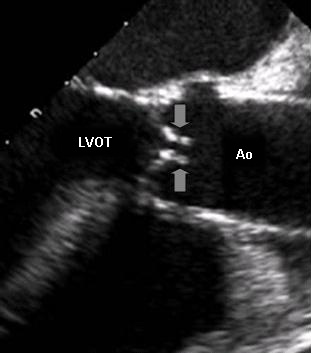
Aortic stenosis
Transesophageal echocardiogram showing the left ventricle outflow tract (LVOT), the aorta (Ao), and nearly immobile leaflets (arrows) of a severely stenotic aortic valve
From the collection of David Liff, MD, Emory University Hospital; used with permission
See this image in context in the following section/s:
Videos
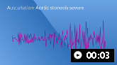 Aortic stenosis (severe)
Auscultation sounds: Aortic stenosis (severe)
 How to perform an ECG: animated demonstration
How to record an ECG. Demonstrates placement of chest and limb electrodes.
Use of this content is subject to our disclaimer