Tests
1st tests to order
ECG
Test
This nonspecific test is performed at baseline and again if there is a change in the patient's rhythm.
Result
atrial fibrillation; left atrial enlargement; right ventricular hypertrophy
CXR
Test
Test ordered at baseline and again if there is a major change in symptoms.[Figure caption and citation for the preceding image starts]: Kerley B lines (horizontal lines in the left lower zone): a radiologic change of pulmonary venous hypertensionFrom Harley HR. Br Heart J. 1961 Jan;23(1):75-87. Used with permission [Citation ends].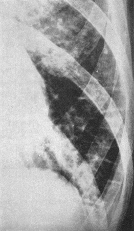
Result
double right heart border indicating an enlarged left atrium; prominent pulmonary artery; Kerley B lines
transthoracic echocardiography
Test
When the typical findings of mitral stenosis are discovered on physical examination, an echocardiogram is the definitive test to confirm the diagnosis and to quantitate the severity of the disease.[22][23][Figure caption and citation for the preceding image starts]: Transthoracic echocardiogram: long-axis view of a rheumatic mitral valve showing classic doming of the leaflets in diastoleFrom Siva A, Shah AM. Heart. 2005 Jan;91(1):e3. Used with permission [Citation ends].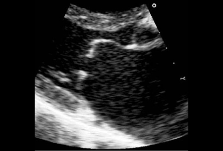 [Figure caption and citation for the preceding image starts]: Short-axis view of left ventricle with flattening of the interventricular septum secondary to right ventricular pressure overloadFrom Siva A, Shah AM. Heart. 2005 Jan;91(1):e3. Used with permission [Citation ends].
Used to measure mitral orifice area and assess pulmonary artery pressure.
Also used to establish suitability of valve anatomy for balloon valvotomy.
Result
hockey stick-shaped mitral deformity
Tests to consider
transesophageal echocardiography (TEE)
Test
Used to examine for left atrial thrombus that might be dislodged during valvotomy.[22][23]
Additionally, TEE may be helpful in assessing mitral annular calcification-related mitral stenosis.
Result
presence of possible left atrial thrombus; calcification of mitral annulus; mitral leaflets are usually normal although in some cases the basal parts of the leaflets may be calcified
cardiac catheterization
Test
Used to calculate valve area.
Only performed if inadequate information is available from echocardiography and surgical correction is planned.
Result
high left atrial pressure, low left ventricular pressure, and low cardiac output
dynamic exercise testing
Test
Exercise response measured by echocardiography or cardiac catheterization.
If pulmonary capillary wedge (PCW) pressure or pulmonary artery pressure increase with exercise, the patient should be considered for valvotomy.
Result
pressures increase with exercise; PCW >25 mmHg or pulmonary systolic pressure >60 mmHg
Use of this content is subject to our disclaimer