Images and videos
Images


Mitral stenosis
Stages of mitral stenosis (rheumatic disease)
Adapted from Otto CM et al. J Am Coll Cardiol 2021:77:e25-e197
See this image in context in the following section/s:
Mitral stenosis
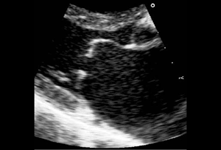
Transthoracic echocardiogram: long-axis view of a rheumatic mitral valve showing classic doming of the leaflets in diastole
From Siva A, Shah AM. Heart. 2005 Jan;91(1):e3. Used with permission
See this image in context in the following section/s:
Mitral stenosis
Gorlin formula. MVA, mitral valve area; CO, cardiac output; HR, heart rate; dfp, diastolic filling period (seconds); p1-p2, mean mitral gradient
See this image in context in the following section/s:
Mitral stenosis
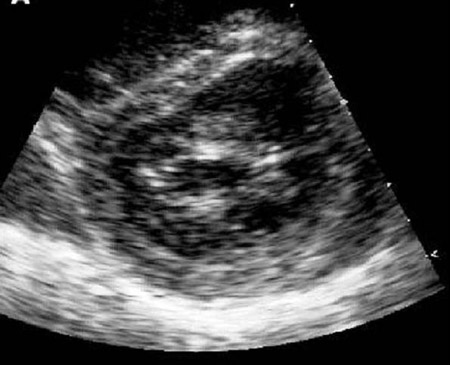
Short-axis view of left ventricle with flattening of the interventricular septum secondary to right ventricular pressure overload
From Siva A, Shah AM. Heart. 2005 Jan;91(1):e3. Used with permission
See this image in context in the following section/s:
Mitral stenosis
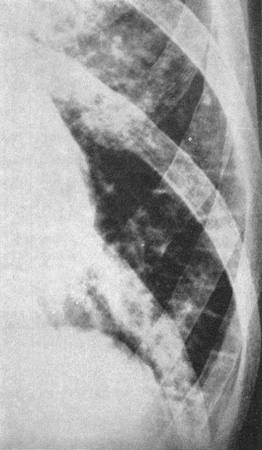
Kerley B lines (horizontal lines in the left lower zone): a radiologic change of pulmonary venous hypertension
From Harley HR. Br Heart J. 1961 Jan;23(1):75-87. Used with permission
See this image in context in the following section/s:
Videos
 Mitral stenosis (severe)
Auscultation sounds: Mitral stenosis (severe)
Use of this content is subject to our disclaimer