Images and videos
Images


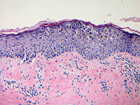

Melanoma
Photomicrograph of melanoma in situ
From the personal collection of Dr Hobart Walling and Dr Brian Swick.
See this image in context in the following section/s:

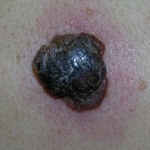
Melanoma
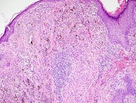
Nodular melanoma
From the personal collection of Dr Hobart Walling and Dr Brian Swick.
See this image in context in the following section/s:

Melanoma
Bluish-white veil of a melanoma
From the personal collection of Dr Hobart Walling and Dr Brian Swick.
See this image in context in the following section/s:

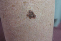
Melanoma
Superficial spreading melanoma
From the personal collection of Dr Hobart Walling and Dr Brian Swick.
See this image in context in the following section/s:

Melanoma
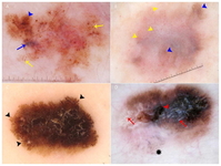
Key dermoscopic features of melanoma: (A) Melanoma presenting with atypical globules and dots of different sizes and shapes (yellow arrows), patches of atypical network (blue arrowhead) and a blue-white veil (blue arrow). (B) Melanoma with diffuse polymorphous vasculature, consisting of serpentine, dotted, and glomerular vessels, can be found throughout the lesion (yellow arrowheads); in addition, patches of atypical network (blue arrowheads) are seen. (C) Superficial spreading melanoma with pseudopods distributed asymmetrically around the lesion (black arrowheads). (D) Melanoma with the regression structure blue-gray peppering (black star); shiny white lines are also seen throughout the entire lesion (red arrows) along with a central blue-white veil (red arrowhead)
Wolner ZJ et al. Enhancing skin cancer diagnosis with dermoscopy. Dermatol Clin. 2017 Oct;35(4):417-37; used with permission
See this image in context in the following section/s:
Melanoma
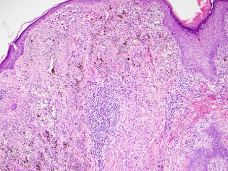
Photomicrograph of Clark level IV invasive melanoma
From the personal collection of Dr Hobart Walling and Dr Brian Swick.
See this image in context in the following section/s:

Melanoma
Fitzpatrick skin type
Created by the BMJ Knowledge Centre
See this image in context in the following section/s:

Melanoma
Dermoscopy: the most important application of dermoscopy is distinguishing melanoma from benign melanocytic lesions
© DermNet New Zealand; used with permission
See this image in context in the following section/s:

Melanoma
Subungual melanoma in situ
From the personal collection of Dr Hobart Walling and Dr Brian Swick.
See this image in context in the following section/s:
Use of this content is subject to our disclaimer