Images and videos
Images


Endometrial cancer
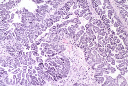
Histologic subtype: clear cell adenocarcinoma (photomicrograph, hematoxylin and eosin stain)
Courtesy of Professor Robert H. Young, Department of Pathology, Massachusetts General Hospital
See this image in context in the following section/s:
Endometrial cancer
Histologic subtype: uterine papillary serous carcinoma with typical small papillae and slit-like spaces (photomicrograph, hematoxylin and eosin stain)
Courtesy of Professor Robert H. Young, Department of Pathology, Massachusetts General Hospital
See this image in context in the following section/s:
Endometrial cancer
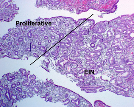
Epithelial in situ neoplasia arising in proliferative endometrium.(photomicrograph, hematoxylin and eosin stain)
From the collection of George Mutter MD, Division of Women's and Perinatal Pathology, Brigham and Women's Hospital, Harvard Medical School
See this image in context in the following section/s:
Endometrial cancer
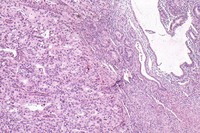
Grade 3 or high-grade endometrioid adenocarcinoma on a background of atrophic endometrium (photomicrograph, hematoxylin and eosin stain)
Courtesy of Professor Robert H. Young, Department of Pathology, Massachusetts General Hospital
See this image in context in the following section/s:
Endometrial cancer
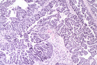
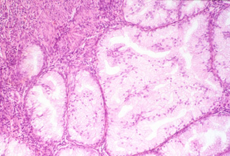
Histologic subtype: endometrioid endometrial adenocarcinoma, the commonest subtype; diagnosed on dilation and curettage in a patient presenting with postmenopausal bleeding (photomicrograph, hematoxylin and eosin stain)
Courtesy of Professor Robert H. Young, Department of Pathology, Massachusetts General Hospital
See this image in context in the following section/s:

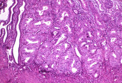
Endometrial cancer
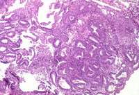
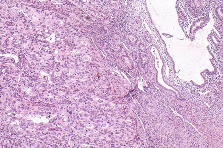
Grade 1 or low-grade endometrioid adenocarcinoma (right) on background of proliferative endometrium (left) (photomicrograph, hematoxylin and eosin stain)
Courtesy of Professor Robert H. Young, Department of Pathology, Massachusetts General Hospital
See this image in context in the following section/s:

Endometrial cancer
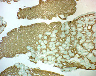
Low-grade adenocarcinoma arising in proliferative endometrium, stained using immunohistochemistry (brown stain) for phosphatase and tensin homolog protein; note the negatively stained (nonbrown) neoplastic glands (normal epithelium usually stains brown)
From the collection of George Mutter MD, Division of Women's and Perinatal Pathology, Brigham and Women's Hospital, Harvard Medical School
See this image in context in the following section/s:
Use of this content is subject to our disclaimer