Approach
Initial evaluation involves taking a detailed history, a physical exam (including a digital rectal exam [DRE]), and initial investigations.
History
A detailed history includes:
A description of the patient's bowel habits
An assessment of the duration and severity of symptoms
Any history of precipitating events, including drug history.
Specific questions concerning bowel habit include: details of stool frequency, stool consistency (may be rated using the Bristol stool form scale [BSFS]), stool size, urge to defecate, degree of straining during defecation, sensation of incomplete evacuation, the need for digital disimpaction of stool, and any history of ignoring a call to stool.
The history should ascertain the onset, severity, and duration of each symptom. Patients frequently define constipation on the basis of excessive straining, a sense of incomplete evacuation, failed or lengthy attempts to defecate, hard stools, and less frequently by the number of stools per week.[1][2][3] The American College of Gastroenterology describes constipation as unsatisfactory defecation characterized by infrequent stools, difficult stool passage, or defecation that is both infrequent and/or difficult.[4]
The patient should also be questioned about the presence of risk factors for constipation. Risk factors include female sex, ages >65 years, a low fiber diet, inadequate fluid or calorie intake, a sedentary lifestyle, and use of any constipation-inducing drugs (e.g., opioids, calcium-channel blockers, antipsychotics, tricyclic antidepressants).[18][30][35] Surgical procedures and childbirth may also be associated with constipation, including any history of abdominal surgery, vaginal delivery, need for episiotomy, or obstetric injury.
Consider opioid-induced constipation in patients who are using opioids.[18]
Bristol stool form scale and stool diaries
A prospective 7-day stool diary can be helpful in documenting stool frequency and consistency, and other symptoms of constipation.[37] Digital stool apps, such as Constipation Stool Diary, have been validated as an alternative to paper stool diaries.[38][39]
Retrospective recall of stool patterns can often be unreliable.[40] A BSFS of ≤3 indicates hard stool.
Type 1 - separate hard lumps, like nuts (hard to pass)
Type 2 - sausage-shaped, but lumpy
Type 3 - like a sausage but with cracks on its surface
Type 4 - like a sausage or snake, smooth and soft
Type 5 - soft blobs with clear-cut edges (passed easily)
Type 6 - fluffy pieces with ragged edges, a mushy stool
Type 7 - watery, no solid pieces (entirely liquid).
Physical examination
Physical exam includes a detailed neurologic exam to evaluate for signs of neuropathy, diabetes, hypothyroidism, gastrointestinal cancer, and neurologic problems. The abdomen is examined for masses or presence of stool in the left or right lower abdominal quadrants. Anorectal inspection looks for skin excoriations, skin tags, anal fissures, or hemorrhoids. Assessment of perineal sensation and anocutaneous reflex by gently stroking the perianal skin with a cotton bud (Q-tip) or blunt needle in all four quadrants will elicit reflex contraction of the external anal sphincter. If this reflex is absent, a neuropathy should be suspected.
A DRE is performed to evaluate for rectal strictures, stool in the rectal vault, heme-positive stool, and rectoceles. If there is a suspicion for a rectocele in a female patient, a vaginal exam can be helpful.[9][41] The anal sphincter tone is assessed, both at rest and during squeeze.
If stool is present, its consistency should be noted and patients should be asked if they were aware of its presence. A lack of awareness of stool in the rectum may suggest rectal hyposensitivity.
Assessing the resting and squeeze tone of the anal sphincter and puborectalis muscle is done by asking the patient to squeeze. The patient should be asked to push and bear down as if to defecate. During this maneuver, the examiner should perceive relaxation of the external anal sphincter and/or the puborectalis muscle, together with perineal descent. A hand placed on the abdomen can gauge the abdominal push effort. An absence of these normal findings should raise the index of suspicion for an evacuation disorder such as dyssynergic defecation.[8]
Alarm features
Alarm features detected during the history or physical exam include:
Rectal bleeding
Heme-positive stool
Weight loss
Obstructive symptoms
Recent onset of symptoms
Rectal prolapse
Change in stool caliber
Age >50 years.
The presence of any of these features indicates a higher risk for a secondary cause for the constipation. Subsequent steps in diagnostic assessment and initial management will depend on the presence or absence of alarm features.[Figure caption and citation for the preceding image starts]: Diagnostic algorithm for constipation. Traditional laxatives include bulk laxatives, osmotic laxatives, stimulant laxatives, and stool softeners. See Management approach section for further details. AXR: abdominal x-rayCreated by BMJ Knowledge Centre from material supplied by Satish Rao, MD and Ashok Attaluri, MD [Citation ends].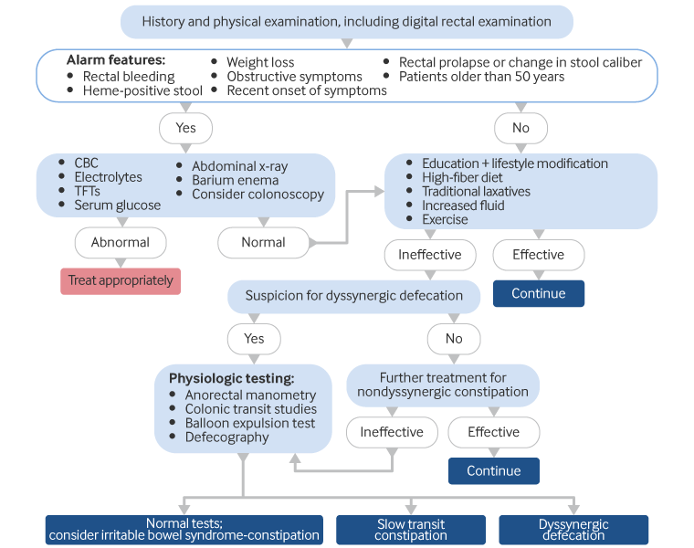
Initial investigations in people with alarm features
A CBC, serum electrolytes (including calcium and magnesium), glucose, and thyroid function studies are performed when there are clinical suspicions of a secondary cause.[9][41] There are no studies demonstrating the utility of performing blood tests in all patients presenting with constipation.
Abdominal x-ray and barium enemas are performed, although their exact specificity and sensitivity in this setting are not known. Abdominal x-ray can be helpful in assessing colonic stool load in obese patients, where physical exam is difficult. Colonoscopy may also be considered. The American Society of Gastrointestinal Endoscopy recommends colonoscopy in patients with constipation only if they have alarm features or iron-deficiency anemia. Patients age >50 years may be considered for colonoscopy if they have not previously had colon cancer screening.[42]
UK guidelines recommend quantitative fecal immunochemical testing (FIT) to guide referral for suspected colorectal cancer in adults with a change in bowel habit.[43] If FIT result is ≥10 micrograms hemoglobin/g feces, refer patients urgently. Referral should not be delayed if there is a strong clinical suspicion (e.g., due to abdominal mass) and FIT is negative or if patients do not return their sample.[43]
If any of these investigations reveal a secondary cause, it should be treated appropriately. However, if all investigations are normal, patients can be managed and investigated further as though they have no alarm features.
Initial management of people with no alarm features
The following are initial treatments, the results of which help to decide on the need for further investigation in people with no alarm features:
Patient education
Advice to eat a high-fiber diet
Advice to drink more fluids
Advice to take regular exercise
Traditional laxatives.
See Management approach.
Abdominal x-ray can be helpful in assessing colonic stool load in obese patients where physical exam is difficult.
Further investigation (people with no alarm features, and people with alarm features with normal initial investigations)
If initial treatment measures are effective, they should be continued. However, if they are ineffective after approximately 6 to 8 weeks, the diagnosis of dyssynergic defecation is considered. Symptoms associated with dyssynergia include:
A sensation of anal blockage
Straining
The use of digital maneuvers to assist defecation.
Patients with constipation and negative evaluations and/or a lack of response to conventional therapy should have physiologic testing. Physiologic testing includes colonic transit studies, anorectal manometry, balloon expulsion studies, and colonic manometry (indicated in selected patients). Although there is no definitive evidence that this affects the outcome of treatment, this approach defines the underlying pathophysiology and facilitates targeted therapy.[4][18][44]
Defecography may also be performed at this stage. Defecography can provide useful information about anatomy, and can help in assessing several parameters of anorectal function (anorectal angle at rest and during straining, perineal descent, anal diameter, indentation of the puborectalis, amount of rectal and rectocele emptying).[18][45] As this procedure involves exposure to high levels of radiation, the clinician should exercise caution in obtaining this test, especially in women with child-bearing potential. Dynamic pelvic magnetic resonance imaging (magnetic resonance defecography) is a newer modality that can simultaneously evaluate global pelvic floor anatomy and dynamic motion.[46][47][48] However, since normative data for both barium and MR defecography are scarce, the distinction between normal and abnormal pathology is challenging at times, and the two modalities should be considered complementary.[18] In one Cochrane review of imaging studies in women with symptoms of obstructed defecation, the pooled sensitivity for a barium study compared with MR defecography for pelvic floor descent was 98% versus 94%, and pooled specificity was 83% versus 79%, respectively.[49]
These further tests may help to diagnose slow-transit constipation or dyssynergic defecation. However, these tests are not widely available and frequently need referral to a tertiary level hospital. If physiologic testing is normal, the diagnosis of irritable bowel syndrome-constipation predominant (IBS-C) may be considered, especially if abdominal pain is one of the predominant symptoms.
Use of this content is subject to our disclaimer