Images and videos
Images


Chronic coronary disease
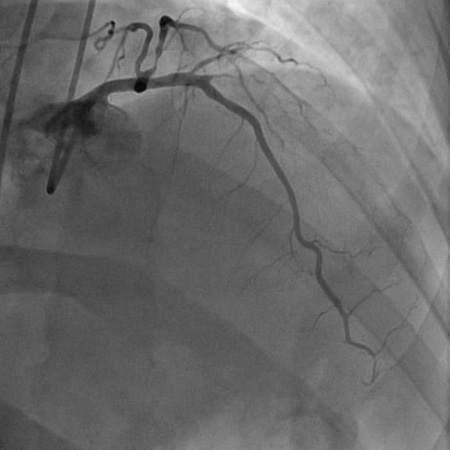
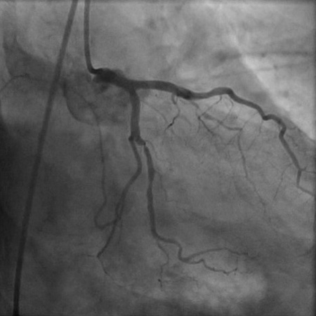
Angiogram (right anterior oblique cranial projection) in a 55-year-old man with a 1-month history of angina on exertion. The image shows a 90% proximal stenosis of obtuse marginal 1 (explaining the patient's lateral ischemia), 90% proximal stenosis of the first diagonal, and 99% subtotal occlusion of the second diagonal (explaining the patient's anterior and anterolateral ischemia)
From the collection of Dr S.D. Fihn; used with permission
See this image in context in the following section/s:
Chronic coronary disease
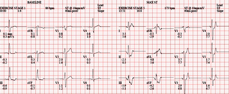
Computerized summary of exercise ECG in a 55-year-old man with a 1-month history of angina on exertion
From the collection of Dr S.D. Fihn; used with permission
See this image in context in the following section/s:
Chronic coronary disease
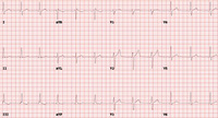
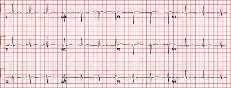
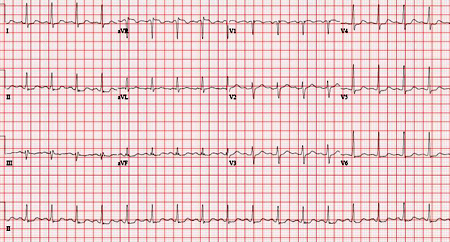
Normal ECG
From the collection of Dr S.D. Fihn; used with permission
See this image in context in the following section/s:
Chronic coronary disease
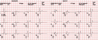
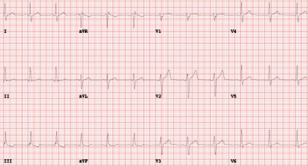
Baseline exercise ECG in a 55-year-old man with a 1-month history of angina on exertion
From the collection of Dr S.D. Fihn; used with permission
See this image in context in the following section/s:
Chronic coronary disease
Sensitivity and specificity of tests for anatomically significant coronary artery disease
Adapted from Knuuti et al. The performance of non-invasive tests to rule-in and rule-out significant coronary artery stenosis in patients with stable angina: a meta-analysis focused on post-test disease probability. Eur Heart J. 2018 Sep 14;39(35):3322-30; used with permission. (CCTA, coronary computed tomography angiography; CI, confidence interval; CMR, stress cardiac magnetic resonance; PET, positron emission tomography; SPECT, single-photon emission computed tomography [exercise stress SPECT with or without dipyridamole or adenosine]; Stress echo, exercise stress echocardiography)
See this image in context in the following section/s:
Chronic coronary disease
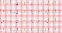
ECG showing nonspecific ST depressions in V5 and V6, which may indicate ischemia. There are nonspecific ST-segment changes in III and aVF
From the collection of Dr S.D. Fihn; used with permission
See this image in context in the following section/s:
Chronic coronary disease
Angiogram (right anterior oblique caudal projection) in a 55-year-old man with a 1-month history of angina on exertion. A 90% proximal stenosis of obtuse marginal 1 is present, explaining the patient's lateral ischemia
From the collection of Dr S.D. Fihn; used with permission
See this image in context in the following section/s:

Chronic coronary disease
Pretest probabilities of obstructive coronary artery disease in symptomatic patients according to age, sex, and nature of symptoms in pooled analysis
Juarez-Orozco et al. Eur Heart J Cardiovasc Imaging. 2019 Nov 1;20(11):1198-207; used with permission
See this image in context in the following section/s:

Chronic coronary disease
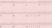
Maximal exercise ECG in a 55-year-old man with a 1-month history of angina on exertion with ST depressions in II, III, aVF diagnostic of ischemia, and normal ST changes in V4-6 (rapid upsloping)
From the collection of Dr S.D. Fihn; used with permission
See this image in context in the following section/s:
Videos
 How to perform an ECG: animated demonstration
How to record an ECG. Demonstrates placement of chest and limb electrodes.
Use of this content is subject to our disclaimer