Approach
Primary care physicians are generally on the front line of care for patients with diabetes-related foot disease. As such, most of the initial evaluation and management is done through primary care clinics. Endocrinologists and other medical specialists may also be involved in the evaluation and management of these patients. An interprofessional approach facilitated by a podiatrist is recommended by the American Diabetes Association for all patients with foot ulcers and high-risk feet.[22]
The main goals of the initial evaluation include:
Identifying the presence of any foot ulcers
Assessing for any clinical symptoms or signs of infection
Assessing for the presence of sensory neuropathy
Documenting pedal pulses
This should be done in all patients with diabetes, even in the absence of any suspicion of diabetes-related foot disease.
An active ulcer immediately requires a greater sense of urgency and should be classified according to the degree of tissue loss, the presence/degree of ischemia, and the presence/degree of infection. The key factors associated with occurrence or recurrence include the presence of sensory neuropathy (loss of protective sensation), the presence of vascular disease, and/or a past history of an ulcer, Charcot neuro-osteoarthropathy, or amputation. These three factors can easily be screened without complex equipment.[21]
See Criteria for guidance on the classification of ulcers, infection and risk stratification, and Screening for the recommended frequency of foot checks.
History
Strong risk factors for diabetes-related foot disease include: sensory neuropathy; peripheral artery disease; previous history of foot ulcer, infection, or partial amputation; cigarette smoking; chronic kidney disease (including end-stage renal disease), and foot deformities, in particular those resulting from Charcot neuro-osteoarthropathy (i.e., midfoot collapse).[22][23] Lack of protective sensation is most often due to diabetes, but can occasionally be due to other causes (e.g., alcohol misuse).
Most patients who develop foot ulcers have at least some degree of sensory neuropathy, which reduces the nociceptive feedback that usually signals an injury sustained during ambulation or via a puncture wound. However, it is common for patients to note the onset of foot pain in a previously insensate area when an infection is present. A history of fever, chills, malaise, or anorexia is suggestive of an infection. A history of claudication or rest pain is suggestive of peripheral artery disease.
Physical exam
The physician should examine the skin integrity of the foot and any muscular deformities in a well-lit room. When the feet are examined in a patient with diabetes, shoes, socks, bandages, and dressings should be removed, and both feet should be examined for the following:[22][38]
Skin: assess integrity, color, and temperature, as well as the presence of ulcers, callus, fungal infection, puncture wounds, gangrene, and pre-ulcerative signs such as hemorrhage or fissures.
Bone and joint deformities such as bunions, hammertoes, prominent metatarsals, and limited joint mobility.
Loss of protective sensation due to peripheral sensory neuropathy: use a 10-g monofilament plus at least one other of pinprick, temperature perception, ankle reflexes, or vibratory perception with a 128-Hz tuning fork or similar device. The monofilament test is considered positive if monofilament is not detected in any of the four tested areas in the forefoot.[15] The Ipswich Touch Test is a suitable alternative for screening and detection of high-risk inpatients requiring foot protection.[24]
Peripheral artery disease (PAD): assess lower-extremity pulses, capillary refill time, rubor on dependency, pallor on elevation, and venous filling time. If there is suspicion of PAD, consider performing Doppler ultrasound with pulse volume recordings, and calculation of the ankle-brachial index and toe systolic blood pressure.
Infection and/or inflammation: edema, localized warmth, and erythema are suggestive of cellulitis, with or without a deep soft-tissue infection (i.e., abscess). Fluctuance is also suggestive of an abscess.
Charcot neuro-osteoarthropathy.
Footwear and signs of poor self-care, such as unwashed feet or improperly cut toenails.
The American Diabetes Association's Comprehensive Diabetic Foot Exam (CDFE) has been condensed into a 3-minute foot exam designed to retain the above key factors while dramatically reducing time for a clinician to perform a thorough assessment.[40]
When examining the feet, pay special attention to weight-bearing areas, as the majority of nonhealing foot ulcers and infections occur in these areas, resulting from repetitive trauma during ambulation on an insensate, sometimes structurally abnormal foot. The majority of nonhealing foot ulcers and foot infections occur in the forefoot, the portion of the foot distal to the tarsometatarsal (Lisfranc) joint. Patients with Charcot neuro-osteoarthropathy with midfoot collapse may develop ulcers and infections in the midfoot that are associated with structural abnormalities there. Heel ulcers occur less frequently in ambulatory patients, and are often due to decubitus pressure in nonambulatory patients debilitated by previous stroke. Leg/calf ulcers (occurring between the knee and the malleoli at the ankle) are generally due to chronic venous insufficiency.
If any signs of infection are found on examination, classify and document the severity as mild, moderate or severe using the International Working Group on the Diabetic Foot (IWGDF)/Infectious Diseases Society of America (IDSA) system.[41] It is worth noting that because of the impaired immune response and abnormal arteriovenous shunting present in the neuropathic foot, clinical signs of infection in patients with diabetes may be more subtle than in patients without diabetes.[Figure caption and citation for the preceding image starts]: Midfoot ulcer in a patient with Charcot neuro-osteoarthropathy (midfoot collapse)From the collection of Dr Neal R. Barshes; used with permission [Citation ends].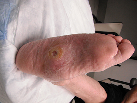 [Figure caption and citation for the preceding image starts]: Uninfected foot ulcer overlying the plantar aspect of the first metatarsophalangeal joint. Note the hyperkeratotic skin (callus) surrounding the wound edgeFrom the collection of Dr Neal R. Barshes; used with permission [Citation ends].
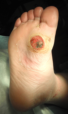 [Figure caption and citation for the preceding image starts]: A foot infection originating from a gangrenous third toe. Note the erythema and fluctuance in the midfoot. An abscess cavity was found tracking under the longitudinal section of macerated skinFrom the collection of Dr Neal R. Barshes; used with permission [Citation ends].
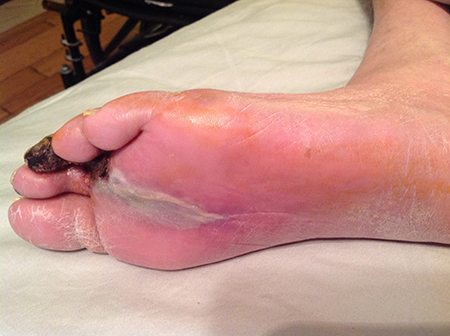
When examining any infected foot ulcer, it is also essential to consider whether there is underlying osteomyelitis, as its presence greatly increases the risk of amputation.[41] Osteomyelitis is particularly likely in wounds that are wide, deep, have been present for many weeks, are located over a bony prominence, show visible bone, or are accompanied by an erythematous, swollen ("sausage") toe.[41]
To assess for osteomyelitis, perform a probe-to-bone test as part of the physical exam, by gently inserting a sterile blunt metal probe into the wound. The test is defined as positive if the user feels a hard, gritty structure (i.e., bone) against the end of the probe. Although its reliability varies by users’ technique and experience, overall the probe-to-bone test is the most useful examination technique for diagnosing osteomyelitis in diabetic foot infections (in conjunction with imaging and serum biomarkers), according to the IWGDF and IDSA. Its sensitivity and specificity are 0.87 and 0.83, respectively, suggesting that a positive result in a high-risk patient supports the diagnosis, whereas a negative result in a low-risk patient helps rule it out.[41]
Pedal pulse examination
Pulse exam is the most accessible modality for evaluating arterial blood flow to the foot; however, even when performed by an experienced physician such as a vascular surgeon, inter-observer agreement is modest. The exam can be further impaired by the foot and ankle edema that is common in the setting of a foot infection. Nevertheless, the ability to palpate normal pedal pulses indicates adequate arterial perfusion to the foot. Absent or weak pulses should prompt referral for evaluation and noninvasive testing in the form of Doppler ultrasound with pulse volume recordings.[38]
Investigations
The diagnosis of diabetes-related foot disease is fundamentally a clinical diagnosis based on thorough history and examination: no blood tests are universally recommended, beyond those which form part of routine diabetes care. However in all patients with a suspected foot infection, initial investigations should include a blood glucose level, and inflammatory biomarkers such as white blood cell count, C-reactive protein, erythrocyte sedimentation rate, or procalcitonin.[41] Do not perform procalcitonin testing without an established, evidence-based protocol.[42] Leukocytosis may suggest the presence of an infection; however, this test has medium sensitivity/specificity.[43] Renal function tests may provide prognostic information; presence of chronic kidney disease increases risk of amputation and all-cause mortality.[44][45] Renal function tests can also be helpful in determining the feasibility of giving iodinated contrast for arterial imaging (if necessary). See Acute kidney injury (Prevention).
When a diabetic foot infection is suspected, samples should be taken for culture to allow selection of appropriate antibiotic therapy.[41] Tissue specimens collected via curettage or biopsy provide culture results with higher specificity and sensitivity than superficial swabs, although are more burdensome to collect.[41] In low-resource settings, a Gram-stain smear may be used as an alternative to culture to visualize the class of causative pathogen.[41] Do not take samples for culture if the wound is not clinically infected. False positive culture results may lead to the unnecessary prescription of antibiotics, which could cause harmful side effects and promote antibiotic resistance.[46]
Consider x-rays if the clinical examination is suggestive of any bone or joint deformities, fractures, osteomyelitis or Charcot neuro-osteoarthropathy.[8] Weight-bearing films should be considered whenever feasible, especially in patients with Charcot neuro-osteoarthropathy.[31][38][47]
Characteristic radiographic features of osteomyelitis include loss of bone cortex, focal loss of trabecular pattern, periosteal reaction, bone sclerosis, and abnormal soft tissue density in the subcutaneous fat suggesting a deep ulcer or sinus tract.[41] Plain x-rays are less sensitive during the acute phase of osteomyelitis, and should be repeated in 2-3 weeks if suspicion is still high after a normal initial x-ray.[38]
Although x-ray remains first line for diagnosing osteomyelitis due to its low cost and widespread availability, further imaging may be required, particularly to differentiate from noninfectious structural changes related to Charcot neuro-osteoarthropathy. Magnetic resonance imaging of the foot is considered the best imaging test for this purpose and is recommended by the IWGDF/IDSA when the initial x-ray is normal and the clinical suspicion of osteomyelitis remains.[41] 18F-fluorodeoxyglucose positron emission tomography (FDG-PET/CT), 99mTc-exametazime Hexa Methyl Propylene Amine Oxime (HMPAO)-labeled white blood cell scintigraphy, or 99mTc-labeled Ubiquicidin (UBI) SPECT/CT single photon emission computed tomography (SPECT/CT) can be considered as alternatives to MRI for diagnosing osteomyelitis.[41]
Making an accurate diagnosis of osteomyelitis in a diabetic foot can be challenging as there is no universally accepted definition or criteria, and low levels of agreement between commonly used diagnostic tests.[41] Osteomyelitis may be present in a patient with diabetes despite normal inflammatory markers, x-rays, or probe-to-bone testing.[8] See Complications.
Peripheral artery disease (PAD)
Noninvasive vascular testing, in the form of Doppler ultrasound with pulse volume recordings and ankle/toe pressures, can aid the diagnosis of peripheral artery disease and should be performed in any patient with a history and examination suggestive of PAD, according to American Diabetes Association guidelines.[22]
The Society for Vascular Surgery (SVS) and the American Podiatric Medical Association go further, advocating universal PAD screening with noninvasive arterial studies for all people with diabetes ages over 50 years.[48] If normal, these should be repeated every 5 years.[22] The American College of Cardiology Foundation and the American Heart Association recommend a resting ankle-brachial index (ABI) be performed in all people with suspected PAD, defined as one or more of the following: nonhealing foot wounds, exertional leg symptoms, age 65 years and older, or 50 years or older with diabetes or a history of smoking.[49]
ABIs should be calculated as part of noninvasive vascular testing but interpreted carefully, as results can be inaccurate in people with diabetes due to arterial calcification.[22] It is important, therefore, not to rule out a diagnosis of PAD in a patient with diabetes based solely on a normal or raised ABI. The ADA recommends measuring toe systolic blood pressure, as these are more accurate than ABI alone: toe pressures <30 mmHg are suggestive of PAD and poor ulcer healing. Individuals with abnormal pulse volume tracings and toe pressures <30 mmHg with foot ulcers should be referred for immediate vascular evaluation.[22]
Joint guidelines from the International Working Group on the Diabetic Foot (IWGDF), European Society for Vascular Surgery (ESVS) and SVS, published in 2023, emphasize that no single test has been found to reliably exclude PAD in patients with a diabetic foot ulcer. They recommend evaluation of pedal Doppler waveforms in combination with ankle systolic pressure, ABI, toe systolic pressure, and toe brachial index (TBI). PAD is less likely if ABI is 0.9 to 1.3, TBI is ≥0.70, and triphasic or biphasic pedal Doppler waveforms are present.[10] Toe waveforms and pressures are preferred in people with diabetes.[10][50]
Angiography is considered to be the best test for diagnosing PAD, and should be performed when considering a revascularization procedure.[10] Angiography can provide detailed anatomic information on the lower limb vasculature including the presence, severity and distribution of hemodynamically significant (i.e., >50%) arterial stenoses or occlusions between the aorta and the foot. Imaging should extend all the way from the aorta to the foot, with detailed imaging of the tibial and pedal vessels in particular.[10] Decisions regarding the need for angiography should generally be made by a vascular specialist. Modalities include computed tomographic angiography, magnetic resonance angiography, and catheter digital subtraction angiography. Of these, catheter digital subtraction angiography is considered the gold standard, according to IWGDF/ESVS/SVS guidelines. However the authors note that each technique has its advantages and disadvantages, and the choice depends on local availability and expertise.[10]
For more information, see Peripheral arterial disease.
Use of this content is subject to our disclaimer