History and exam
Key diagnostic factors
common
history of diabetes mellitus
Present in the majority of patients presenting with a foot ulcer or foot infection, and in all patients with diabetes-related foot disease.
presence of risk factors
Key risk factors include sensory neuropathy, peripheral arterial disease, previous history of foot ulcer, foot deformity, limited foot and/or ankle joint mobility, previous history of major amputation, and end-stage renal disease requiring renal replacement therapy.
foot ulcer
A diabetic foot ulcer is defined as a break in the skin of the foot in a person with diabetes that includes as a minimum the epidermis and part of the dermis.[1] Most occur in the forefoot, the portion of the foot distal to the tarsometatarsal (Lisfranc) joint.
Patients with Charcot neuro-osteoarthropathy leading to midfoot collapse may develop ulcers and infections in the midfoot that are associated with structural abnormalities there.
Heel ulcers are often due to decubitus pressure in nonambulatory patients debilitated by previous stroke.[Figure caption and citation for the preceding image starts]: Midfoot ulcer in a patient with Charcot neuro-osteoarthropathy (midfoot collapse)From the collection of Dr Neal R. Barshes; used with permission [Citation ends].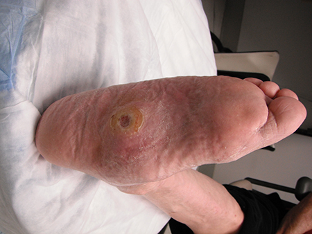 [Figure caption and citation for the preceding image starts]: Uninfected foot ulcer overlying the plantar aspect of the first metatarsophalangeal joint. Note the hyperkeratotic skin (callus) surrounding the wound edgeFrom the collection of Dr Neal R. Barshes; used with permission [Citation ends].
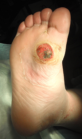
foot pain
Most patients who develop foot ulcers have at least some degree of sensory neuropathy. Sensory neuropathy blunts or obviates the nociceptive feedback that usually signals an injury sustained during ambulation or via a puncture wound. However, it is common for patients to note the onset of foot pain in a previously insensate area when an infection is present.
loss of protective sensation
A sign of peripheral neuropathy, characterized by an inability to sense light pressure. Assessed using a 10-g monofilament plus at least one other of pinprick, temperature perception, ankle reflexes, or vibratory perception with a 128-Hz tuning fork or similar device.[22]
foot deformity
Foot deformities can cause alteration in pressure distribution, predisposing the skin to traumatic ulceration.
uncommon
fever or chills
Suggests infection.
Other diagnostic factors
common
malaise
Suggests infection.
anorexia
Suggests infection.
foot erythema
Suggests cellulitis with or without deep soft-tissue infection (i.e., abscess). [Figure caption and citation for the preceding image starts]: A foot infection originating from a gangrenous third toe. Note the erythema and fluctuance in the midfoot. An abscess cavity was found tracking under the longitudinal section of macerated skinFrom the collection of Dr Neal R. Barshes; used with permission [Citation ends].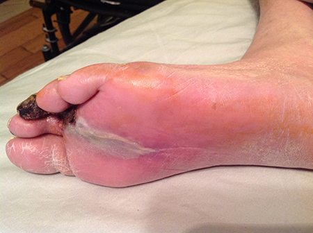
However, in the absence of an open ulcer the diagnosis of acute Charcot neuro-osteoarthropathy in a patient with peripheral sensory neuropathy should also be considered.
edema of foot, ankle, or calf
Suggests infection.
absent pedal pulses
Consistent with the presence of peripheral artery disease.
Ability to palpate normal pedal pulses indicates adequate arterial perfusion to the foot. Absent or weak pulses should prompt referral for evaluation and noninvasive testing in a vascular specialty clinic.
Augmenting the exam with a handheld continuous-wave Doppler probe provides additional information when properly performed and interpreted. However, while monophasic signals do suggest significant peripheral artery disease, biphasic signals do not exclude significant peripheral artery disease.
uncommon
fluctuance
Suggests a deep soft-tissue infection (i.e., abscess). [Figure caption and citation for the preceding image starts]: A foot infection originating from a gangrenous third toe. Note the erythema and fluctuance in the midfoot. An abscess cavity was found tracking under the longitudinal section of macerated skinFrom the collection of Dr Neal R. Barshes; used with permission [Citation ends].
Risk factors
strong
sensory neuropathy
Aside from a previous history of ulcer or amputation, sensory neuropathy is the single most influential factor associated with the risk of foot ulcers (and subsequent infection or limb loss).[11][21] It blunts or obviates the nociceptive feedback that usually signals an injury sustained during ambulation or via a puncture wound. Every patient with diabetes should have a foot assessment at least annually that includes a sensory examination using a 10-g monofilament plus at least one other of pinprick, temperature perception, ankle reflexes or vibratory perception with a 128-Hz tuning fork or similar device.[22][23] The Ipswich Touch Test has also been shown to be a simple, reproducible method that may be practical for many settings.[24][25]
previous history of foot ulcer
Approximately 40% of patients with a previous foot ulcer have a recurrence within 1 year of ulcer healing, with almost 60% experiencing a recurrence within 3 years and 65% within 5 years.[6]
previous history of amputation
peripheral artery disease (PAD)
Diabetes increases the risk of developing peripheral artery disease (PAD), and the presence of PAD increases the risk of foot ulcer development.[10] The prevalence of PAD in people with diabetes is 20% to 28%, rising to 50% among those with established diabetic foot ulcers.[10] In the overwhelming majority of these cases, the chronic impairment in arterial blood flow to the foot is not the direct cause of the foot ulcer. Rather, PAD of varying severity impairs the normal inflammatory response to foot trauma and, therefore, mainly represents an impediment to healing once an epithelial defect is already present. Every patient with diabetes should be checked for PAD at least annually: as a minimum this should include assessment of peripheral pulses, capillary refill time, rubor on dependency, pallor on elevation, and venous filling time.[22][23] If examination or history are suggestive of PAD, patients should be referred for noninvasive arterial studies and/or further vascular assessment as appropriate.[22]
end-stage renal disease
Although the precise mechanism is not known, end-stage renal disease has a significant impact on both the risk of developing and the ability to heal foot ulcers. Patients with end-stage renal disease, including anyone on renal replacement therapy, are at high risk of developing foot ulcers.[23] Renal insufficiency is also associated with the development of PAD and peripheral neuropathy.[27][28]
foot deformities
Structural foot deformities pose an ulceration risk by leading to improper distribution of pressure across the foot during ambulation. The presence of any foot deformity, in combination with sensory neuropathy or peripheral arterial disease, is considered a moderate risk factor for ulceration in the 2023 International Working Group on the Diabetic Foot (IWGDF) risk stratification system. A foot deformity is defined as alterations or deviations from the normal shape or size of the foot, such as hammer toes, mallet toes, claw toes, hallux valgus, prominent metatarsal heads, pes cavus, pes planus, pes equinus, or results of Charcot neuro-osteoarthropathy, trauma, amputations, other foot surgery or other causes.[23]
Midfoot deformity as a result of Charcot neuro-osteoarthropathy (i.e., midfoot collapse) is uncommon, but when present it can represent a significant challenge. Midfoot ulcers associated with deformity resulting from Charcot neuro-osteoarthropathy are difficult to offload and heal. Osteomyelitis in the midfoot is more difficult to address with surgery without jeopardizing the foot stability.[Figure caption and citation for the preceding image starts]: Midfoot ulcer in a patient with Charcot neuro-osteoarthropathy (midfoot collapse)From the collection of Dr Neal R. Barshes; used with permission [Citation ends].
limited foot and/or ankle joint mobility
Joint immobility in patients with diabetes mellitus is thought to be the result of deposition of advanced glycation end-products.
Stiffness of the Achilles tendon and/or gastrocnemius muscle may reduce ankle dorsiflexion, thereby increasing pressure in the forefoot during the push-off phase of gait. An inability to passively dorsiflex the ankle past neutral (i.e., to passively achieve an angle of <90° between the plantar foot and the calf) is considered abnormal.[29]
There is some evidence that addressing ankle equinus via orthopedic/podiatric lengthening procedures may help the healing of forefoot ulcers and may reduce their recurrence.[30]
weak
visual impairment
In addition to hindering the ability to visually inspect one's feet, visual impairment in the setting of diabetes mellitus is often a marker of microvascular complications.
poor glucose control
There is a very clear causal relationship between poor glucose control and the development of sensory neuropathy (among other microvascular complications). However, after adjusting for the presence or absence of sensory neuropathy, glucose control itself has a weaker association with the development of foot ulcers.[11]
Charcot’s neuro-osteoarthropathy
The presence of Charcot’s neuro-osteoarthropathy predisposes to bone and joint deformities, which are themselves risk factors for development of foot ulceration and infection. However, there is currently insufficient evidence to support the inclusion of Charcot’s neuro-osteoarthropathy as an independent risk factor for these outcomes by itself, according to the IWGDF.[23][31]
obesity
Use of this content is subject to our disclaimer