Criteria
A diabetic foot ulcer is a break in the skin that includes as a minimum the epidermis and part of the dermis and which occurs below/distal to the malleoli in a person with diabetes.[1] By history and clinical examination, diabetic foot ulcers can be classified as neuropathic, neuro-ischaemic (a combination of neuropathy and ischaemia), or ischaemic.[36] The majority of foot ulcers are purely neuropathic or neuro-ischaemic. Only a small percentage are purely ischaemic: these tend to be painful and follow from minor trauma.[36]
SINBAD system for classification of diabetic foot ulcers
The SINBAD (Site, Ischaemia, Neuropathy, Bacterial Infection, Area, and Depth) scoring system is recommended by the IWGDF for assessing and classifying diabetic foot ulcers, as well as for audit, benchmarking, and communication between healthcare professionals.[7][45] It is well validated and simple to use, and contains the majority of prognostic clinical features that predict outcome including likelihood of amputation. SINBAD is also recommended by NICE and is the system used by the UK National Diabetes Foot Care Audit.[9][46]
SINBAD uses a scoring system with a maximum of 6 points. A score of 3 or more is associated with an increased time to healing and greater risk of eventual failure to heal.[45] When using the SINBAD system, for communication between healthcare professionals, clinicians should describe the individual variables rather than a total score.[7]
Site
Forefoot (0 points)
Midfoot and hindfoot (1 point)
Ischaemia
Pedal blood flow intact: at least one palpable pulse (0 points)
Clinical evidence of reduced pedal flow (1 point)
Neuropathy
Protective sensation intact (0 points)
Protective sensation lost (1 point)
Bacterial infection
None (0 points)
Present (1 point)
Area
Ulcer <1 cm² (0 points)
Ulcer ≥1 cm² (1 point)
Depth
Ulcer confined to skin and subcutaneous tissue (0 points)
Ulcer reaching muscle, tendon, or deeper (1 point)
WIfI scoring
The WIfI (Wound, Ischaemia, Foot Infection) scoring system is also widely used for classifying and describing diabetic ulcers, and it is recommended for this purpose by the American Heart Association on the grounds that it also helps determine amputation risk and aids clinical decision-making.[18] The IWGDF also endorses the WIfI system as a valid alternative to SINBAD for classifying and describing diabetic ulcers, provided there is sufficient expertise and resources to use it.[7] As with SINBAD, it is recommended that clinicians report the individual variables that make up the WIfI system, rather than giving a total score.
The WIfI system can also be used in two other clinical situations: to stratify the likelihood of healing and amputation risk in patients with diabetes, peripheral arterial disease, and a foot ulcer or gangrene; and to describe infected ulcers (although the IWGDF/Infectious Diseases Society of America [IDSA] classification is recommended as first-choice for the latter scenario).[7]
Wound (W):
0: no ulcer or gangrene
1: mild - small, shallow ulcer(s) on distal leg or foot; no exposed bone (unless limited to distal phalanx); no gangrene
2: moderate - deeper ulcer with exposed bone, joint, or tendon; generally not involving the heel; shallow heel ulcer without calcaneal involvement; gangrenous changes limited to digits
3: severe - extensive, deep ulcer involving forefoot and/or midfoot; deep, full thickness heel ulcer and/or calcaneal involvement; extensive gangrene involving forefoot and/or midfoot; full thickness heel necrosis and/or calcaneal involvement
Ischaemia (I):
0: ankle-brachial index (ABI) ≥0.80; ankle systolic pressure >100 mmHg; toe pressure (TP)/transcutaneous oximetry (TcPO₂) ≥60 mmHg
1: mild - ABI 0.6 to 0.79; ankle systolic pressure 70-100 mmHg; TP/TcPO₂ 40-59 mmHg
2: moderate - ABI 0.4 to 0.59; ankle systolic pressure 50-70 mmHg; TP/TcPO₂ 30-39 mmHg
3: severe - ABI ≤0.39; ankle systolic pressure <50 mmHg; TP/TcPO₂ <30 mmHg
Foot infection (fI):
0: no symptoms or signs of infection
1: mild - infection present, as defined by the presence of at least two of the following:
Local swelling or induration
Erythema >0.5 cm to ≤2 cm around ulcer
Local tenderness or pain
Local warmth
Purulent discharge
2: moderate - local infection (as described above) with erythema >2 cm, or involving structures deeper than skin and subcutaneous tissues (e.g., abscess, osteomyelitis, septic arthritis, fasciitis); no systemic inflammatory response signs
3: severe (limb and/or life-threatening) - local infection (as described above) with signs of systemic inflammatory response syndrome as manifested by at least two of the following:
Temperature >38°C (>100.4°F) or <36°C (<96.8°F)
Heart rate >90 bpm
Respiratory rate >20 breaths/minute or PaCO₂ <32 mmHg
WBC count >12 × 10⁹ cells/L (12,000/microlitre) (leukocytosis) or <4 × 10⁹ cells/L (4000/microlitre) (leukopenia); or a normal WBC count with >10% immature (band) forms
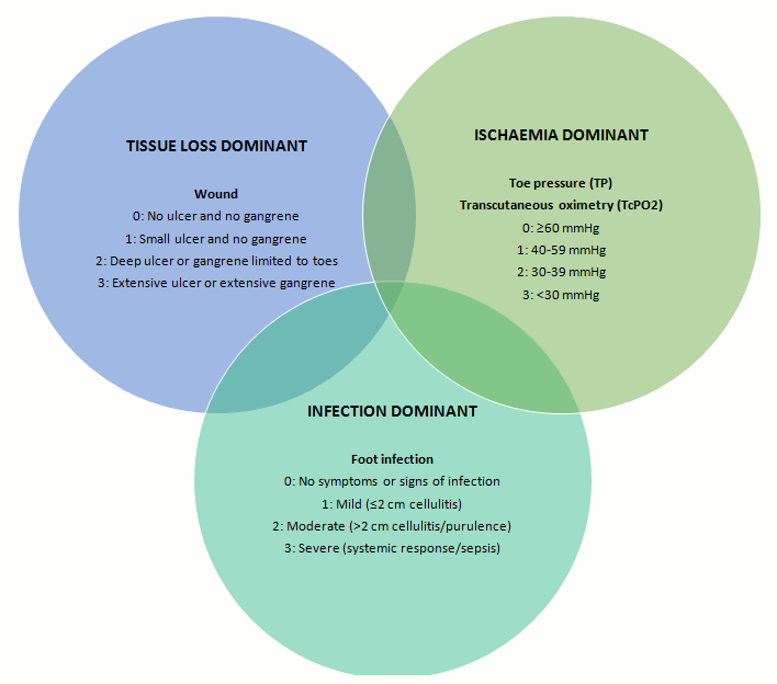
A simple Venn diagram has been designed to assist clinicians in using the WIfI system, to determine which of the three factors (wound, ischaemia, or infection) is dominant.
[Figure caption and citation for the preceding image starts]: Diabetic foot problems can be related to the presence of a wound, ischaemia, or infection (WIfI). Which of these parameters is dominant can vary, and a flexible long-term management approach is needed. The Venn diagram shows intersecting rings of dominance for these three parameters, with gradings listed for each. The shaded areas represent combinations of these parameters of dominanceFrom the collection of Dr David G. Armstrong and Dr Joseph L. Mills Sr; used with permission [Citation ends].
Foot infections
According to NICE and the IWGDF/IDSA, a diabetic foot infection is defined by the presence of at least two of the following:[9][40]
Local swelling or induration
Erythema (>0.5 cm around the wound)
Local tenderness or pain
Local warmth
Purulent discharge
IWGDF/IDSA infection classification
In a person with diabetes and an infected foot ulcer, the IWGDF and NICE recommend the IWGDF/IDSA infection classification as the first choice to characterise the infection and guide management.[9][40] The WIfI system, described above, can be used as an alternative for this purpose.
IWGDF/ISDA classification consists of three grades of severity for diabetic foot infection. It was originally developed as part of the PEDIS classification for research purposes and is used as a guideline for management, in particular to identify which patients require hospital admission for intravenous antibiotics. It is a strong predictor of the need for hospitalisation.[48] It has also been validated for risk of both major and minor amputation.[49][50]
Infection severity[40]
1: Uninfected
No systemic or local symptoms or signs of infection
2: Mild
Presence of ≥2 of the following: local swelling or induration, erythema 0.5 to <2.0 cm around the wound, local tenderness or pain, local increased warmth, or purulent discharge (exclude other causes of inflammatory response, such as trauma, gout, acute Charcot neuro-osteoarthropathy, fracture, thrombosis, and venous stasis).
3: Moderate: Add 'O' for any infection involving bone (osteomyelitis)
Infection (as for mild severity above) with no systemic manifestations and involving erythema extending ≥2 cm from the wound margin, and/or tissue deeper than skin and subcutaneous tissues (e.g., tendon, muscle, joint, and bone). Add 'O' for any infection involving bone (osteomyelitis).
4: Severe: Add 'O' for any infection involving bone (osteomyelitis)
Any foot infection with associated manifestations of systemic inflammatory response syndrome, as manifested by ≥2 of the following: temperature >38°C or <36°C, heart rate >90 beats/minute, respiratory rate >20 breaths/minute or PaCO₂ < 4.3 kPa (32 mmHg), WBC count >12 × 10⁹ cells/L (12,000/microlitre) (leukocytosis) or <4 × 10⁹ cells/L (4000/microlitre) (leukopenia); or a normal WBC count with >10% immature (band) forms.
Use of this content is subject to our disclaimer