Differentials
Atopic dermatitis
SIGNS / SYMPTOMS
History of atopy and lack of lesions with active inflammatory border with central clearing. Lesions in flexural folds of neck, arms, and legs. Chronic and relapsing, often since childhood.
INVESTIGATIONS
Diagnosis is clinical. Potassium hydroxide (KOH) microscopy is infrequently done, and rarely fungal culture or biopsy may be undertaken to exclude diagnosis of dermatophyte infection.
Dyshidrotic dermatitis
SIGNS / SYMPTOMS
Sudden eruption of pruritic, small intradermal vesicles on hands and feet. There may be history of contact irritants: for example, nickel.
INVESTIGATIONS
Diagnosis is clinical. KOH microscopy is infrequently done and rarely fungal culture or biopsy may be undertaken to exclude diagnosis of dermatophyte infection.
Lichen simplex chronicus
SIGNS / SYMPTOMS
Leather-like, hyperpigmented patches that are pruritic and chronically rubbed, producing a leathery change in affected skin.
INVESTIGATIONS
Diagnosis is clinical. KOH microscopy is infrequently done, and rarely fungal culture or biopsy may be undertaken to exclude diagnosis of dermatophyte infection.
Psoriasis
SIGNS / SYMPTOMS
Family history of psoriasis, presence of characteristic silvery plaque and lesions located on extensor surface of elbows and knees will aid in diagnosis. Psoriasis limited to the nails can be difficult to distinguish, particularly if limited to toenails. In psoriasis, subungual hyperkeratosis is usually white silver in color. Pitting of nails may be mistaken for tinea unguium. Clinical differentiation will usually suffice for tinea capitis, unguium, or corporis. [Figure caption and citation for the preceding image starts]: Annular lesion on the elbow, with a silvery scale. No central clearing. Microscopic exam with potassium hydroxide revealed no fungal elements. Despite the resemblance to tinea corporis, there was a similar lesion on the extensor surface of both knees and a family history that together confirmed the diagnosis of psoriasisDepartment of Dermatology Medical University of South Carolina; used with permission [Citation ends].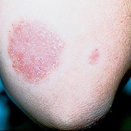
INVESTIGATIONS
KOH microscopy is infrequently done, and rarely fungal culture or biopsy may be undertaken to exclude dermatophyte infection.
Trichotillomania
SIGNS / SYMPTOMS
History of obsessive habit of twisting hair with fingers. Usually no inflammatory changes in scalp, and broken hairs are of different lengths.
INVESTIGATIONS
Clinical diagnosis. Trichoscopy shows broken hairs of different length.
Traction alopecia
SIGNS / SYMPTOMS
History of tightly braided hair styles. Broken hairs with patchy alopecia. Usually no scaling and inflammation of scalp
INVESTIGATIONS
Clinical diagnosis.
Alopecia areata
SIGNS / SYMPTOMS
Complete rather than patchy hair loss.
INVESTIGATIONS
Diagnosis is clinical. Trichoscopy shows "exclamation mark" hair and other types of broken hairs.
Erythema chronicum migrans
SIGNS / SYMPTOMS
Rapidly enlarging and reddening, single or multiple bull's eye lesions on trunk. History of tick exposure or associated symptoms of Lyme disease.
INVESTIGATIONS
Occasionally, KOH microscopy is required to distinguish this from tinea corporis. Antibody titers or skin biopsy for diagnosis of Lyme disease.
Pityriasis versicolor
SIGNS / SYMPTOMS
Hypopigmented truncal lesions in dark-skinned individuals, darker than normal color with scaling in light-skinned individuals; commonly, below neck level.
INVESTIGATIONS
Clinical differentiation usually sufficient; occasionally KOH microscopy is required to distinguish this from tinea corporis.
Ultraviolet light from Wood lamp shows a pale yellow-white fluorescence.
Pseudofolliculitis barbae
SIGNS / SYMPTOMS
Hyperpigmented nodules in beard area with incurving hairs in patients with dark skin.
INVESTIGATIONS
Clinical diagnosis.
Seborrheic dermatitis
SIGNS / SYMPTOMS
Greasy and scaly area in scalp (with scaling but no hair loss, i.e., dandruff) and erythema in the nasolabial folds and occasionally central chest.
INVESTIGATIONS
Clinical diagnosis.
Acne rosacea
SIGNS / SYMPTOMS
Acneiform eruption with erythema and easy blushing in nasal and malar area.
INVESTIGATIONS
Clinical diagnosis.
Discoid lupus erythematosus
SIGNS / SYMPTOMS
Malar rash, sun sensitivity.
INVESTIGATIONS
Skin biopsy is confirmatory.
Contact dermatitis
SIGNS / SYMPTOMS
Pattern of eruption, intense pruritus, erythema, and occasional vesicular eruption.
INVESTIGATIONS
Clinical diagnosis.
Candidal intertrigo
SIGNS / SYMPTOMS
Usually uniformly red without central clearing or sparing of scrotum; satellite lesions.[Figure caption and citation for the preceding image starts]: Infant presenting with rash formerly known as moniliasis, now called candidiasis, caused by Candida spp.Public Health Image Library, CDC [Citation ends].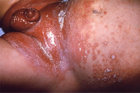
INVESTIGATIONS
Clinical differentiation usually sufficient from tinea cruris; responds to all topical therapies recommended for tinea cruris.
Erythrasma
SIGNS / SYMPTOMS
Uniformly brown and scaly, with no active edge; groin or axillae.
INVESTIGATIONS
Fluoresces a brilliant coral red under Wood lamp.
Friction blisters of feet
SIGNS / SYMPTOMS
Absence of interdigital maceration or moccasin pattern of scaling; bulla primarily at points of contact with ill-fitting footwear; acute in onset.
INVESTIGATIONS
Clinical diagnosis.
Onychogryphosis
SIGNS / SYMPTOMS
Thickened and deviated toenails resembling a ram's horn. Easily misdiagnosed as tinea unguium, this change occurs in older adults where vascular disease and diabetes may play a role. Recurrent trauma (such as that caused by ill-fitting shoes) may play a role.
INVESTIGATIONS
KOH microscopy of nail scrapings, fungal culture, or nail biopsy may be necessary to distinguish from tinea unguium; both conditions may co-exist.
Use of this content is subject to our disclaimer