History and exam
Key diagnostic factors
common
scaling scalp lesions
Scalp lesions in tinea capitis may be irregular or well-demarcated.[Figure caption and citation for the preceding image starts]: Tinea capitisFrom the collection of Professor Antonella Tosti; used with permission [Citation ends].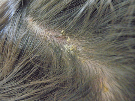 [Figure caption and citation for the preceding image starts]: Tinea capitis in a child with Fitzpatrick type VI skin with the typical appearance of fine scale and brown hair, which may be visualised as black dotsGzzz, Wikimedia Commmons CC-BY-SA-4.0 [Citation ends].
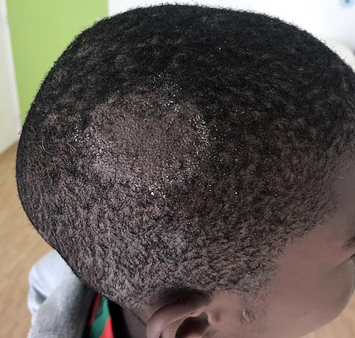
patchy alopecia
Typical in tinea capitis.
erythematous, scaling skin lesions with central clearing
Lesions of face, trunk, extremities, hands, and groin (tinea faciale, tinea corporis, tinea manuum, tinea cruris) reveal a characteristic pattern of inflammation, termed an active border. The inflammatory response is characterized by a greater degree of redness and scaling at the edge of the lesion, with occasional blister formation. Central clearing of the lesion is often present and distinguishes dermatophytoses from other papulosquamous eruptions such as psoriasis or lichen planus, in which there is a uniform inflammatory response throughout the skin lesion.
[Figure caption and citation for the preceding image starts]: Tinea corporis of the axilla. Central clearing with an active border of inflammation noted. Satellite lesion is presentDepartment of Dermatology Medical University of South Carolina; used with permission [Citation ends].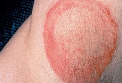 [Figure caption and citation for the preceding image starts]: Annular lesion on the elbow, with a silvery scale. No central clearing. Microscopic exam with potassium hydroxide revealed no fungal elements. Despite the resemblance to tinea corporis, there was a similar lesion on the extensor surface of both knees and a family history that together confirmed the diagnosis of psoriasisDepartment of Dermatology Medical University of South Carolina; used with permission [Citation ends].
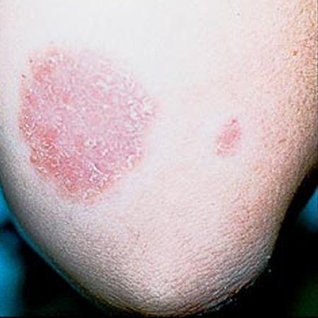
erythematous, scaling rash with follicular pustules in beard or mustache
Typical of tinea barbae, which involves the skin and coarse hairs of the beard and mustache area; occurs in adult men and hirsute women. [Figure caption and citation for the preceding image starts]: Tinea barbae. Note the pustules in the follicles, redness, and scalingDepartment of Dermatology Medical University of South Carolina; used with permission [Citation ends].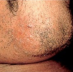
erythematous, annular patches on face
Occurs in tinea facialis. Often these red areas may be indistinct, particularly on darkly pigmented skin, and lesions may have little or no scaling or raised edges. As a result of this subtle appearance, this dermatophytosis is sometimes known as tinea incognito.
vesicles and scaling of hands
May suggest fingernail involvement with tinea manuum.
fissuring, maceration, and scaling in the interdigital spaces of the fourth and fifth toes
The interdigital form of tinea pedis is most common.
chronically, scaly, hyperkeratotic plantar skin with erythema of the soles, heels, and sides of the feet
Tinea pedis, or athlete's foot, may present as a moccasin-like distribution pattern.
folliculitis with nodules
Majocchi granuloma is a foreign-body granuloma caused by dermatophytes, resulting in folliculitis with papules and pustules. Granulomatous nodules are typically in a distinct location, often the lower two-thirds of the leg in women who shave their legs. [Figure caption and citation for the preceding image starts]: Majocchi granulomaFrom the collection of Professor Antonella Tosti; used with permission [Citation ends].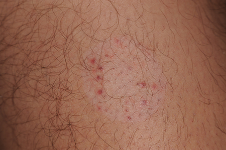
Other diagnostic factors
common
thickened nail with subungual hyperkeratosis, onycholysis, and white-yellow to brown discoloration
Represents distal-lateral subungual onychomycosis.
[Figure caption and citation for the preceding image starts]: Distal lateral subungual onychomycosisFrom the collection of Professor Antonella Tosti; used with permission [Citation ends]. [Figure caption and citation for the preceding image starts]: Vesiculobullous form of tinea pedis and onychomycosisFrom the collection of Professor Antonella Tosti; used with permission [Citation ends].
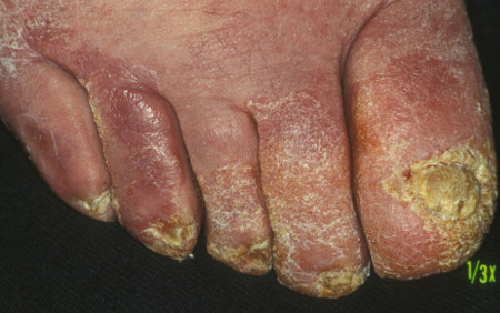
small, white speckled patches on the surface of the nail plate with crumbling nail
Typical of white superficial onychomycosis.
lymphadenopathy
Cervical and occipital lymphadenopathy may be prominent in tinea capitis
uncommon
black-dot alopecia
Swollen hairs may fracture a few millimeters from the scalp, causing this effect.
milky white nail plate
Typical of endonyx onychomycosis.
area of leukonychia in the proximal nail plate
The nail plate becomes white proximally and remains normal distally in proximal subungual onychomycosis. Proximal subungual onychomycosis may be a sign of immunocompromise, such as HIV infection.
Risk factors
strong
exposure to infected people, animals, or soil
Spread by contact is well documented.[11][12]
The skin's barrier mechanisms of protection may fail, leading to trauma, irritation, or maceration of the skin. In tinea barbae the usual cause is a zoophilic organism; therefore, farm workers are most often affected.
Exposure to infected people may occur due to sharing of hats, combs, or hairbrushes, or use of public areas such as swimming pools.[10]
exposure to fomites, including hat, combs, hairbrushes, and upholstery
Indirect transmission of dermatophytes can also occur from fomites.[1]
chronic topical or oral corticosteroid use
Loss of local or systemic immunity is a predisposing factor. With inhibition of immune protection, and/or failure of the skin's protective barrier mechanisms, cutaneous dermatophyte infection may occur.
HIV
Tinea infections, in general, are common.
diabetes mellitus and other metabolic disorders
occlusive clothing
Especially associated with tinea pedis and tinea cruris.
Occlusion of the skin with nonporous materials can interfere with the skin's barrier function to infection by increasing local temperature and hydration levels.[9]
hot, humid weather
Particularly associated with tinea pedis and tinea cruris.
The skin's barrier mechanisms of protection may fail, which can lead to trauma, irritation, or maceration of the skin.
obesity
Especially associated with tinea pedis and tinea cruris.
hyperhidrosis
Especially associated with tinea pedis and tinea cruris.
frequenting public bathing areas while barefoot
deformities of the feet
Associated with tinea pedis and tinea unguium due to occluded and traumatized areas of skin.
recurrent trauma to the skin
Associated with tinea infection.
weak
contact sports
Sports including wrestling, rugby, and football have all been identified as predisposing factors for tinea corporis.[14]
atopic dermatitis
Associated with tinea corporis. Some experts believe a genetic predisposition exists to these infections.
positive family history
For distal subungual onychomycosis caused by infection with Trichophyton rubrum, a family pedigrees study showed a familial pattern indicative of autosomal-dominant inheritance.[5]
peripheral vascular disease
Associated with tinea unguium due to reduced circulation and reduced ability to resist establishment of infection.[15]
Use of this content is subject to our disclaimer