Images and videos
Images


Sarcoidosis
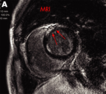
A) Gadolinium-enhanced MRI scan of the heart (short axis) showing delayed enhancement in the anteroseptal myocardium
Adapted from https://casereports.bmj.com/content/2009/bcr.2006.070805.full
See this image in context in the following section/s:
Sarcoidosis
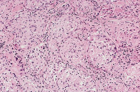
Photomicrograph showing well-formed granulomas typical for sarcoidosis
From the collection of Dr M.P. Muthiah; used with permission
See this image in context in the following section/s:

Sarcoidosis
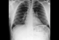
Bilateral hilar adenopathy
From the collection of Dr M.P. Muthiah; used with permission
See this image in context in the following section/s:
Sarcoidosis
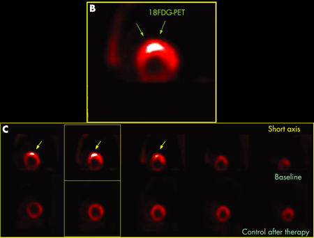
B) 18F-FDG PET scan of the heart (short axis) showing focal uptake in the anteroseptal wall corresponding to granulomatous inflammation. (C) 18F-FDG PET scan of the heart (short axis) from the base to the apex (from left to the right) showing focal uptake in the anteroseptal wall at baseline (upper series) and disappearance of the uptake after treatment (lower series)
Adapted from https://casereports.bmj.com/content/2009/bcr.2006.070805.full
See this image in context in the following section/s:
Use of this content is subject to our disclaimer