Tests
1st tests to order
duplex ultrasound of the leg
Test
If there is a clinical suspicion of popliteal cyst, duplex ultrasound should be obtained as the first diagnostic test.[19] The role of duplex ultrasound is to identify whether there is a cyst and to evaluate for other pathologies such as deep vein thrombosis, hematoma, popliteal artery aneurysm, popliteal vein aneurysm, or soft tissue mass. [Figure caption and citation for the preceding image starts]: Anatomical dissection of the posteromedial knee capsule. The weak area (A) is identified between the 2 expansions of the semimembranosus muscle (B), the oblique popliteal ligament (C), and the expansion over the sheath of the popliteus muscle (D). The semitendinosus muscle (E) and popliteus muscle (F) are also indicatedAdapted from Labropoulos N, Shifrin DA, Paxinos O. New insights into the development of popliteal cysts. Br J Surg. 2004;91:1313-1318; used with permission [Citation ends].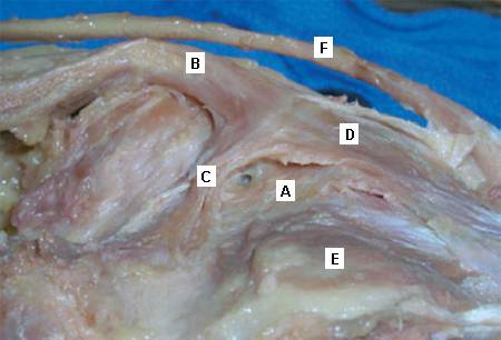
If there is no identified pathology, MRI or CT of the limb should be considered to evaluate for other musculoskeletal or joint pathology.
Result
cystic mass in the posterior-medial popliteal fossa
Tests to consider
MRI of the leg
Test
May identify underlying treatable knee pathology (e.g., arthritis, meniscal tear).
Preferred to CT if ultrasound is negative, because of greater yield.
Result
cystic mass in the posterior-medial popliteal fossa. Uniform hypointense signal on T2 weighted images
CT scan of the leg
Test
May identify underlying treatable knee pathology (e.g., arthritis, meniscal tear), but MRI is the preferred test, with greater yield.
Should be limited to patients who have contraindications to MRI.
Result
cystic mass in the posterior-medial popliteal fossa
Use of this content is subject to our disclaimer