Images and videos
Images


Retinal vein occlusion
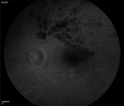
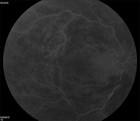
Fluorescein angiogram, left eye; branch retinal vein occlusion; delayed drainage of blocked vein superotemporally
From the personal library of Dr Aziz Khanifar
See this image in context in the following section/s:
Retinal vein occlusion
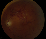
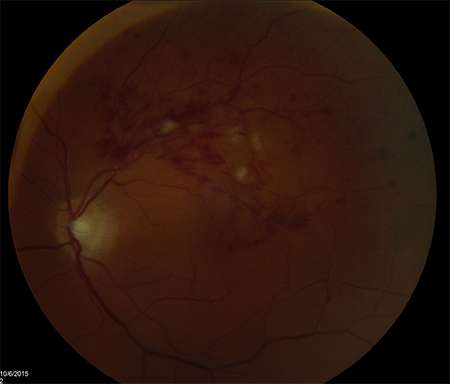
Colour photograph, left eye; branch retinal vein occlusion; multiple intra-retinal images in quadrant of blocked vein
From the personal library of Dr Aziz Khanifar
See this image in context in the following section/s:
Retinal vein occlusion
Fluorescein angiogram, right eye; central retinal vein occlusion; delayed drainage of veins in each quadrant
From the personal library of Dr Aziz Khanifar
See this image in context in the following section/s:

Retinal vein occlusion
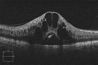
Optical coherence tomogram, right eye; central retinal vein occlusion; large cystoid thickening throughout macula
From the personal library of Dr Aziz Khanifar
See this image in context in the following section/s:

Retinal vein occlusion
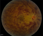
Colour photograph, right eye; central retinal vein occlusion; multiple intra-retinal haemorrhages in each quadrant
From the personal library of Dr Aziz Khanifar
See this image in context in the following section/s:
Use of this content is subject to our disclaimer