History and exam
Key diagnostic factors
common
presence of risk factors
Common risk factors include atherosclerosis, systemic hypertension, diabetes mellitus, history of smoking, cardiovascular disease, glaucoma, increased body mass index at 20 years of age, increased serum alpha-2 globulin, and short axial length.
sudden, painless vision loss
Whole visual field in CRVO, single quadrant in BRVO, altitudinal in hemiretinal vein occlusion (HRVO).
optic nerve head oedema
Present in acute and sub-acute CRVO.
intra-retinal haemorrhage
Present in region of retina affected by RVO: all 4 quadrants in CRVO , 1 quadrant in BRVO, superior or inferior hemisphere in HRVO.[Figure caption and citation for the preceding image starts]: Optical coherence tomogram, right eye; central retinal vein occlusion; large cystoid thickening throughout maculaFrom the personal library of Dr Aziz Khanifar [Citation ends].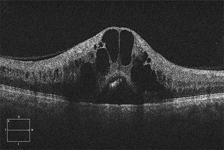 [Figure caption and citation for the preceding image starts]: Colour photograph, right eye; central retinal vein occlusion; multiple intra-retinal haemorrhages in each quadrantFrom the personal library of Dr Aziz Khanifar [Citation ends].
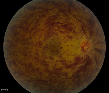
venous tortuosity and dilation
Present in region of retina affected by RVO: all 4 quadrants in CRVO, 1 quadrant in BRVO, superior or inferior hemisphere in HRVO.
uncommon
collateral vessel formation
Can be found in region of optic nerve in chronic CRVO or crossing the horizontal raphe in chronic BRVO.
Other diagnostic factors
common
age >65 years
71%of patients affected by central retinal vein occlusion (CRVO) and 77% of patients affected by branch retinal vein occlusion (BRVO) are >65 years of age.[6]
neovascularisation
Can be present in iris, angle, or retina as a complication of RVO.
vitreous haemorrhage
Associated with neovascularisation, a complication of RVO.
macular oedema
Present as a complication in RVO.
uncommon
floaters
Associated with vitreous haemorrhage, a complication of RVO.
painful, red eye
Associated with neovascular glaucoma, a complication of RVO.
visual acuity
relative afferent pupillary defect
elevated intra-ocular pressure
Intra-ocular pressure may be elevated in patients with glaucoma, which is a risk factor for RVO.[22]
Risk factors
strong
atherosclerosis
Leads to contraction of the common adventitia, turbulent blood flow, endothelial cell injury, and thrombotic venous occlusion.[6]
systemic hypertension
diabetes mellitus
Risk factor for atherosclerosis; associated with increased incidence of RVO in older patients.[6]
hyperlipidaemia
Risk factor for atherosclerosis; incidence of hyperlipidaemia is higher in patients with RVO.[16]
history of smoking
Risk factor for atherosclerosis; associated with increased incidence of RVO in older patients.[6]
cardiovascular disease
Associated with increased incidence of RVO in older patients.
glaucoma
Associated with increased incidence of RVO in older patients.
increased body mass index at 20 years of age
Higher incidence of RVO in patients who had an increased body mass index (>25 g per square metre) as young adults.
increased serum alpha-2 globulin
Higher incidence of RVO in patients with an increased serum alpha-2 globulin (>1.0 g/dl).
short axial length
Axial length usually measured by ultrasound in A-scan mode. However, there is no fixed threshold for short length.[3]
weak
activated protein C resistance
hypercoagulable state
Use of this content is subject to our disclaimer