Images and videos
Images
Acute cervical spine trauma in adults
Canadian C-spine rule. A&E, accident and emergency department; GCS, Glasgow Coma Scale; MVC, Motor vehicle collision. ✝
Adapted from Stiell IG, et al. The Canadian C-spine rule for radiography in alert and stable trauma patients. JAMA. 2001 17;286(15):1841-8.
See this image in context in the following section/s:
Acute cervical spine trauma in adults
Common fracture patterns with severe cervical spine trauma. Top row: cervical burst fracture at C5 level. Top left: axial CT image showing a fracture of C5 vertebral body. Top right: mid-sagittal T2-weighted MRI showing retropulsion of the body of C5 with spinal cord compression, T2-weighted signal changes within the spinal cord, and T2-weighted signal changes within the posterior ligamentous complex indicating disruption of these ligaments. Bottom row: fracture dislocation C6-C7 level. Bottom left: axial CT through C6/C7 facet level. Bottom right: T2-weighted mid-sagittal MRI demonstrating spinal cord compression and T2-weighted signal change within the spinal cord
From the personal collection of Michael G. Fehlings.
See this image in context in the following section/s:

Acute cervical spine trauma in adults
Common fracture patterns with severe cervical spine trauma. Top: axial CT image showing a cervical burst fracture at C5 level. Bottom: axial CT showing fracture dislocation at C6-C7 level.
From the personal collection of Michael G. Fehlings
See this image in context in the following section/s:
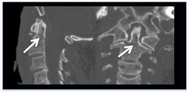
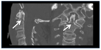
Acute cervical spine trauma in adults
CT reconstruction demonstrating undisplaced odontoid fracture
From the personal collection of Michael G. Fehlings
See this image in context in the following section/s:
Use of this content is subject to our disclaimer