Recommendations
Urgent
Use the Advanced Trauma Life Support protocol to assess and stabilise any patient with suspected trauma. Start with a rapid primary survey using a <C>ABCDE approach:[13][24]
Catastrophic haemorrhage
Airway with in-line spinal immobilisation
Breathing
Circulation
Disability (neurological)
Exposure and environment.
In patients with suspected cervical spine injury carry out and maintain (until the spine is cleared) full in-line spinal immobilisation if the patient has:[13]
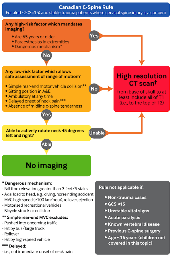
A high-risk factor for cervical spine injury by the Canadian C-spine rule (see flowchart below)
A low-risk factor for cervical spine injury by the Canadian C-spine rule (see flowchart below) AND the patient is unable to actively rotate their neck 45 degrees to both left and right.
Seek urgent advice from a neurosurgeon or spinal surgeon for any patient with a cervical spine injury confirmed on imaging to ensure prompt management.[13]
Assess pain regularly.[13]
Use intravenous morphine as the first-line analgesic.
If intravenous access has not been established, consider diamorphine or ketamine via the intranasal route.
Consider ketamine in analgesic doses as a second-line agent.
Do not use the following medications in the acute phase in patients with a spinal cord injury:[13]
Methylprednisolone
Nimodipine
Naloxone
Medications to prevent neuropathic pain from developing in the chronic stage.
Key Recommendations
[Figure caption and citation for the preceding image starts]: Canadian C-spine rule. A&E, accident and emergency department; GCS, Glasgow Coma Scale; MVC, Motor vehicle collision. ✝Adapted from Stiell IG, et al. The Canadian C-spine rule for radiography in alert and stable trauma patients. JAMA. 2001 17;286(15):1841-8. [Citation ends].
The UK’s National Institute for Health and Care Excellence recommends CT scanning first line in adults who are identified by the Canadian C-spine rule as requiring imaging.[14] When immobilising the spine, manually stabilise the head with the spine in-line using the following stepwise approach:[13]
Fit an appropriately sized, semi-rigid collar unless contraindicated by:
A compromised airway
Known spinal deformities, such as ankylosing spondylitis (in which case keep the spine in the patient's current position)
Reassess the airway after applying the collar
Secure the patient with head blocks and tape.
Discharge patients who have either no indications for a CT scan (as identified by the Canadian C-spine rule) or a normal CT scan, if they are able to rotate their neck 45 degrees to both left and right and do not have severe neck pain (≥7/10 severity).[13]
Advise patients to immediately return to the accident and emergency department if they develop any new neurological symptoms or signs.[33]
Use the Advanced Trauma Life Support protocol to assess and stabilise any patient with suspected trauma. Start with a rapid primary survey using a <C>ABCDE approach:[13][24]
Catastrophic haemorrhage
Airway with in-line spinal immobilisation
Breathing
Circulation
Disability (neurological)
Exposure and environment.
Full in-line spinal immobilisation
Carry out and maintain (until the spine is cleared) full in-line spinal immobilisation if the patient has:[13]
A high-risk factor for cervical spine injury by the Canadian C-spine rule
A low-risk factor for cervical spine injury by the Canadian C-spine rule AND the patient is unable to actively rotate their neck 45 degrees to both left and right
Do not carry out or maintain full in-line spinal immobilisation if the patient:[13]
Has low-risk factors for cervical spine injury by the Canadian C-spine rule AND
Is pain free AND
Is able to actively rotate their neck 45 degrees to both left and right.
Additional injury to the spinal cord can be prevented with timely spinal immobilisation. Prompt detection of these injuries at initial trauma evaluation is imperative. Identifying and assessing C-spine injury during the primary and secondary surveys can be challenging, as patients often present with a decreased level of consciousness, due to concurrent head injury, sedative and analgesic drug, or endotracheal intubation.[8][34] Clinical decision rules in these circumstances are helpful tools for proper management.[8][27][34]
[Figure caption and citation for the preceding image starts]: Canadian C-spine rule. A&E, accident and emergency department; GCS, Glasgow Coma Scale; MVC, Motor vehicle collision. ✝Adapted from Stiell IG, et al. The Canadian C-spine rule for radiography in alert and stable trauma patients. JAMA. 2001 17;286(15):1841-8. [Citation ends].
The UK’s National Institute for Health and Care Excellence recommends CT scanning first line in adults who are identified by the Canadian C-spine rule as requiring imaging[14]When immobilising the spine, manually stabilise the head with the spine in-line using the following stepwise approach.[13]
Fit an appropriately sized, semi-rigid collar unless contraindicated by:
A compromised airway
Known spinal deformities, such as ankylosing spondylitis (in these patients keep the spine in the patient's current position)
Reassess the airway after applying the collar
Secure the patient with head blocks and tape.
Practical tip
Do not attempt to put a collar on a patient who is holding their neck in a fixed position (e.g., a person with ankylosing spondylitis). You may need to allow patients who are uncooperative, agitated, or distressed to assume a position they find comfortable for manual in-line mobilisation.
Practical tip
Cervical spine immobilisation is associated with adverse effects (e.g., raised intracranial pressure, pain, pressure sores). You should remove the collar as soon as is safe and feasible (i.e., as soon as the cervical spine has been cleared by the CT scan, if no abnormalities have been identified).
For patients with suspected thoracic or lumbosacral spine injury see our topic Thoracolumbar spine trauma.
Seek urgent advice from a neurosurgeon or spinal surgeon for any patient with a cervical spine injury confirmed on imaging to ensure prompt management.[13]
Analgesia
Assess pain regularly (using the same pain assessment scale as used in the pre-hospital setting, if this is known).[13] The choice of scale depends on the patient’s age, developmental stage, and cognitive function.[13]
Offer medications to control pain in the acute phase in patients with a spinal injury:[13]
Use intravenous morphine as the first-line analgesic, adjusting the dose as needed to maintain adequate pain relief
If intravenous access has not been established, consider diamorphine or ketamine via the intranasal route (follow local protocols)
Consider intravenous ketamine, given in analgesic doses, as a second-line option.
Practical tip
Do not use the following medications, aimed at providing neuroprotection and prevention of secondary deterioration, in the acute stage in patients with a spinal cord injury:[13]
Methylprednisolone
Nimodipine
Naloxone.
Do not use medications in the acute stage in patients with a spinal cord injury to prevent neuropathic pain from developing in the chronic stage.[13]
Offer oral analgesics to patients with a simple neck sprain injury (i.e., injury to the neck where there has been no demonstrable bony injury or unstable ligamentous injury) or whiplash.
Start with paracetamol. If pain relief is inadequate, replace paracetamol with a nonsteroidal anti-inflammatory drug (NSAID) such as ibuprofen.
Give a weak opioid (e.g., codeine, tramadol) to patients who cannot take an NSAID.
The UK Medicines and Healthcare products Regulatory Agency (MHRA) advises that opioids should be used with caution as there is an increased risk of tolerance, dependence, and addiction, especially with prolonged use (longer than 3 months). The dose should be tapered slowly at the end of treatment to minimise the risk of withdrawal reactions.[35]
As well as providing pain relief in the acute phase after injury, in practice, analgesia is given to enable early mobilisation.
The evidence to support or refute the use of conservative treatments (e.g., rest and wearing a neck collar, physiotherapy, acupuncture, hot and cold packs) in the acute setting remains insufficient.[36]
Discharge with advice
Discharge patients who have either no indications for a CT scan (as identified by the Canadian C-spine rule) or a normal CT scan, if they are able to rotate their neck 45 degrees to both left and right and do not have severe neck pain (≥7/10 severity).[13]
Advise patients to immediately return to the accident and emergency department if they develop any new neurological symptoms or signs.[33]
Use of this content is subject to our disclaimer