Images and videos
Images
Acute cervical spine trauma in adults
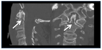
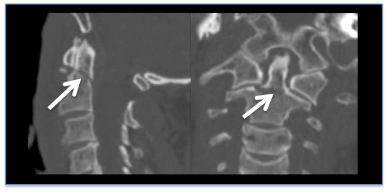
CT reconstruction demonstrating undisplaced odontoid fracture
From the personal collection of Michael G. Fehlings
See this image in context in the following section/s:
Acute cervical spine trauma in adults
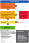
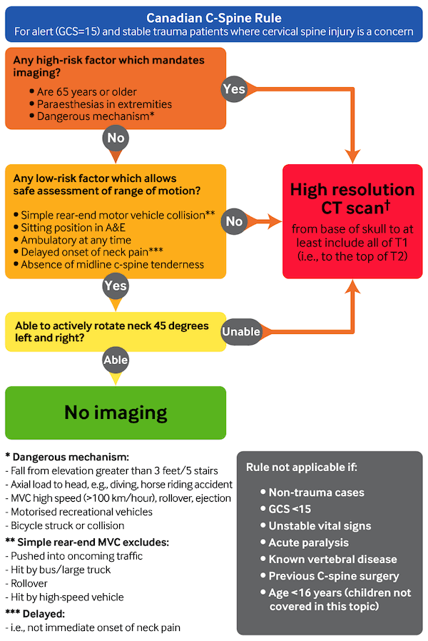
Canadian C-spine rule. A&E, accident and emergency department; GCS, Glasgow Coma Scale; MVC, Motor vehicle collision. ✝
Adapted from Stiell IG, et al. The Canadian C-spine rule for radiography in alert and stable trauma patients. JAMA. 2001 17;286(15):1841-8.
See this image in context in the following section/s:
Acute cervical spine trauma in adults
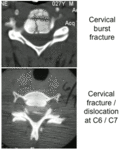
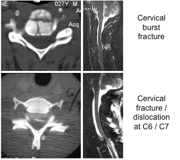
Common fracture patterns with severe cervical spine trauma. Top row: cervical burst fracture at C5 level. Top left: axial CT image showing a fracture of C5 vertebral body. Top right: mid-sagittal T2-weighted MRI showing retropulsion of the body of C5 with spinal cord compression, T2-weighted signal changes within the spinal cord, and T2-weighted signal changes within the posterior ligamentous complex indicating disruption of these ligaments. Bottom row: fracture dislocation C6-C7 level. Bottom left: axial CT through C6/C7 facet level. Bottom right: T2-weighted mid-sagittal MRI demonstrating spinal cord compression and T2-weighted signal change within the spinal cord
From the personal collection of Michael G. Fehlings.
See this image in context in the following section/s:

Acute cervical spine trauma in adults
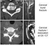
Common fracture patterns with severe cervical spine trauma. Top: axial CT image showing a cervical burst fracture at C5 level. Bottom: axial CT showing fracture dislocation at C6-C7 level.
From the personal collection of Michael G. Fehlings
See this image in context in the following section/s:
Use of this content is subject to our disclaimer