History and exam
Key diagnostic factors
common
presence of risk factors
Key risk factors include adolescent and young adult ages, high ambient temperature and humidity, greasy skin, hyperhidrosis, participation in athletics, and systemic corticosteroid or other immunosuppressant use.
lack of pruritus or pain
PV is most commonly asymptomatic. However, pruritus rarely occurs, and its presence does not exclude the diagnosis of PV.[1]
dyspigmentation
Altered normal skin colour is the primary physical examination finding. These lesions vary in colour, as implied by the name versicolor, from hypopigmented shades of white to hyperpigmented shades of pink, tan, brown, red, or black.[13] Dyspigmentation may be particularly common in people with darker skin, can take months to resolve after successful treatment of infection, and can be psychologically burdensome for some patients.[4][5][Figure caption and citation for the preceding image starts]: Hyperpigmented pityriasis versicolorFrom the collection of Brian L. Swick, MD [Citation ends].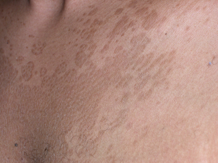 [Figure caption and citation for the preceding image starts]: Hypopigmented pityriasis versicolorFrom the collection of Brian L. Swick, MD [Citation ends].
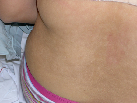 Hypopigmentation is twice as common as hyperpigmentation.[14]
macules or patches
Flat macules becoming confluent to form patches are the most common presentation of PV. Only very rarely is the eruption elevated forming papules or plaques.[1]
seborrhoeic distribution
Distribution of the eruption parallels the density of the sebaceous glands. Commonly affected areas include chest, back, neck, and proximal arms.[1]
fine overlying scale
Pityriasiform scale is common in hyperpigmented lesions of PV but is less common in depigmented lesions.[20]
Other diagnostic factors
uncommon
yellow fluorescence under Wood lamp examination
Lesions may fluoresce a characteristic bright yellow or gold colour under Wood lamp examination. However, Wood lamp examination is positive in only one third of cases, most likely due to most infections being secondary to non-fluorescent species of Malassezia.[13] Only M furfur produces the indole compounds that fluoresce under Wood light.[2][13]
Risk factors
strong
high ambient temperature and humidity
PV is most common in tropical regions and during the summer months in temperate climates when temperature and humidity are highest.[7] As the organism is a lipophilic yeast, requiring exogenous fatty acids for growth, it can thrive and convert from the yeast form to the pathological mycelial form during times of greatest heat, when host sweating and sebum production are at their highest.[7] Even in tropical locations, the disease is most common during months of greatest heat. In one study in Madras, India, PV was most common during May and October, during high ambient temperature.[24]
adolescent and young adult age
Because of the lipid requirements of Malassezia, the yeasts are most commonly found during the years when sebum production is highest and are rarely found on the skin of pre-pubescent children or older people.[7] Although most adults have the yeast on their skin, conversion of the organism from the yeast to the pathological mycelial form requires additional factors such as, in the case of adolescents or young adults, hormonal changes or increased sebum secretion.[1][7]
hyperhidrosis
Because Malassezia yeasts are lipophilic organisms, requiring exogenous fatty acids for growth, hyperhidrosis provides increased host sebum production, allowing the organism to thrive and be converted to its pathological mycelial form.[7]
systemic corticosteroid or other immunosuppressant use
participation in athletics
greasy skin
PV infection has been associated with oily, greasy skin.[25]
weak
family history of PV
Hereditary factors may play a role in susceptibility to PV infection due to the absence of conjugal PV cases and the common appearance of the disease in >1 related family member.[7] The distribution of the disease in first-, second-, and third-degree relatives suggests that susceptibility to PV infection is due to multifactorial inheritance.[26][27]
malnutrition
PV infection has been associated with malnutrition in paediatric cases.[28]
use of oral contraceptives
PV has been associated with use of oral contraceptives.[29]
HIV infection and other immunosuppressive conditions
The prevalence of PV is increased in people with HIV infection and other immunosuppressive conditions. However, the clinical presentation of PV in HIV-positive patients does not differ clinically from PV seen in HIV-negative patients.[30]
use of occlusive ointments or creams
Application to the skin may exacerbate the tendency to develop PV in those prone to the disease.[7]
Use of this content is subject to our disclaimer