Case history
Case history #1
A 20-year-old healthy white man on a basketball team presents with a several-month history of an asymptomatic eruption on the trunk and shoulders. He recalls having a similar eruption several years ago in the summer. On examination, there are slightly scaly, coalescing, fawn-coloured macules and patches on the neck, shoulders, and upper back and chest. There is no evidence of excoriation, erythema, induration, or central clearing.
Case history #2
A 45-year-old Hispanic woman with a medical history of renal failure necessitating renal transplant presents with asymptomatic areas on her shoulders, back, and chest that do not tan. Her current immunosuppressive regimen consists of prednisolone (prednisone) and azathioprine. On examination, she has scaly, hypopigmented, coalescing macules and patches on the neck, shoulders, chest, back, and abdomen. There is no evidence of excoriation, erythema, induration, or central clearing.
Other presentations
The lesions of PV vary in colour (hence versicolor) from hypopigmented and white (PV alba) to hyperpigmented shades of pink, salmon, brown, red (PV rubra) or, rarely, black (PV nigra).[2] PV is generally thought to cause hypopigmented lesions in dark skin and hyperpigmented lesions in light skin. However, one study could find no correlation between pigmentary variation in PV and age, sex, duration, symptoms, or background skin colour in darker skin.[3] Dyspigmentation may be particularly common in people with darker skin.[4][5] PV may rarely be pruritic.[1] Commonly affected areas include the chest, back, shoulders, and neck.[2] Facial involvement is uncommon and is usually seen in tropical climates or in children in temperate climates.[2][6] Less-frequent sites of involvement include the scalp, legs, groin, or within a radiotherapy field.[1] PV is most common in the tropics and in the summer, and in adolescents and young adults.[7] Participation in athletics, pregnancy, hyperhidrosis, malnutrition, immunosuppression, use of oral contraceptives, and family history of PV are associated with the disease.[7][Figure caption and citation for the preceding image starts]: Hypopigmented pityriasis versicolorFrom the collection of Brian L. Swick, MD [Citation ends].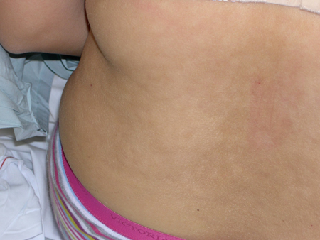 [Figure caption and citation for the preceding image starts]: Pityriasis versicolor in a light-skinned personFrom the collection of Brian L. Swick, MD [Citation ends].
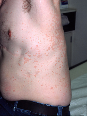 [Figure caption and citation for the preceding image starts]: Hyperpigmented pityriasis versicolorFrom the collection of Brian L. Swick, MD [Citation ends].
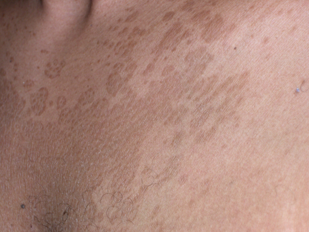
Use of this content is subject to our disclaimer